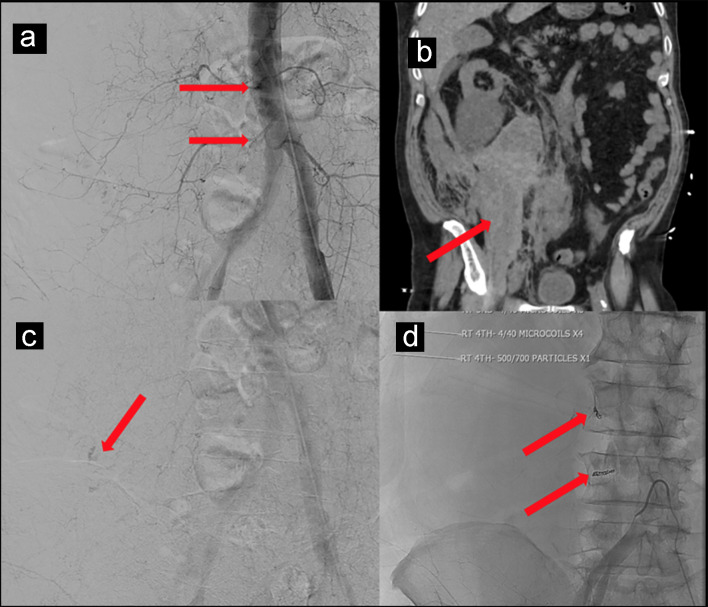
Figure 1.
(a, b) CT scan of the abdomen and pelvis without contrast showing hemorrhage along entire length of right psoas muscle measuring 10 × 17 × 24 cm. (c) Subtle extravasation identified from branches of the right L3 and L4 lumbar arteries. (d) Four 4 mm in diameter by 40 mm in length microcoils placed to control bleeding.

