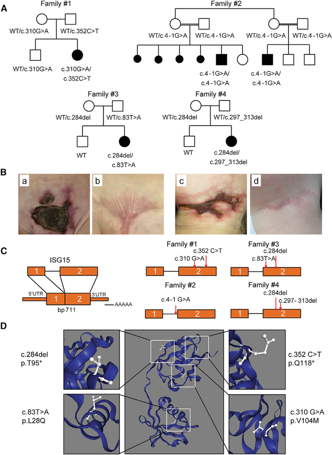
Figure 1. Identification of Five Patients and Six Different Mutations of ISG15.
(A) Familial segregations of the ISG15 alleles. Family 1 includes one affected child (P1) carrying compound heterozygous variants (c.310G>A and c.352C>T) and one unaffected sibling. Family 2 includes two affected children (P2 and P3) with a splice-site variant (c.4–1G>A). Family 3 includes one affected child (P4) with compound heterozygous variants (c.83T>A and c.284del). Family 4 includes one affected child (P5) with compound heterozygous variants (c.284del and c.297_313del). The corresponding genotypes are indicated.
(B) Skin lesions from P1 (a and b) and P4 (c and d).
(C) Schematic localization of the ISG15 variants in the genomic DNA, indicated by the red arrows.
(D) Predicted localization of the four amino acid substitutions in the three-dimensional (3D) model of ISG15.

