
Fig. 3: Conditional PDK4 deletion results in a marked shift in myocardial substrate utilization, decreased DNA damage, and increased proliferation in adult cardiomyocytes.
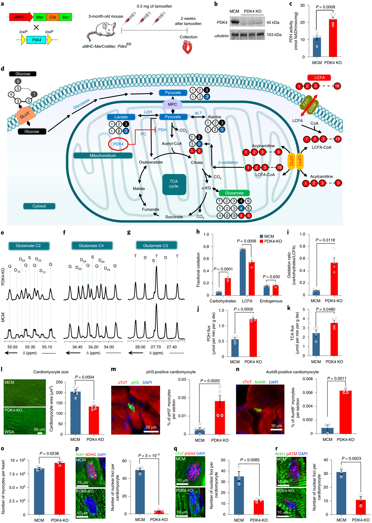
a, Schematic view of the inducible knockout αMHC-MerCreMer cross with PDK4f/f . b, Western Blot for the PDK4 protein shows noticeable depletion in the PDK4 protein expression in the PDK4 KO group compared to the control αMHC-MerCreMer (MCM) hearts. The experiment was repeated twice, with similar results. c, Pyruvate Dehydrogenase (PDH) activity (n=5 biologically independent mice per group). d, A scheme showing the generation of glutamate multiplets from glucose, lactate-pyruvate, and LCFA. Note that 13C labeling for glucose, lactate-pyruvate, and LCFA is indicated as black, blue and red balls, respectively. e, Glutamate C-2 (55.35 ppm) f, Glutamate C-4 (34.20 ppm) and g, Glutamate C-3 (27.60 ppm) spectra. The letters S, D, T, and Q refer to a singlet, doublet (with the relevant J-coupled spins), triplet (a degenerate doublet of doublets) or quartet (or doublet of doublets), respectively. h, Fractional oxidation (n=4 biologically independent mice per group). i, Carbohydrates to LCFA oxidation ratios (n=4 biologically independent mice per group). j, PDH flux and k. TCA flux (n=4 biologically independent mice per group). l, WGA staining shows a significant decrease in cardiomyocyte cell size measurement in PDK4 KO mice compared to the control (n=5 biologically independent mice per group). m, Anti-pH3 and anti-cTnT co-immunostaining shows a significant increase in the cardiomyocyte mitosis marker in cardiomyocyte-specific PDK4 KO mice (n=5 biologically independent mice per group). n, Anti-Aurora B kinase and anti-cTnT co-immunostaining shows a significant increase in cardiomyocyte cytokinesis marker in PDK4 KO compared to the control group (n=3 biologically independent mice per group). o, Quantification of total number of isolated cardiomyocytes by collagenase digestion showing a significant increase in PDK4 KO group at 4 weeks after the first tamoxifen injection (n=3 biologically independent mice per group). Data in p, q and r represent the co-immunostaining with anti-8-hydroxyguanosine (8OHG), anti-γH2AX and anti-pATM antibodies, respectively. There are significant decreases in oxidative DNA damage and the DNA damage response pathway in cardiomyocytes from PDK4 KO hearts (n=3 biologically independent mice per group). Data are presented as the mean±s.e.m. Statistical analysis was performed with two-tailed Student’s t-test: NS, not significant. Actn1, Alpha actinin-1, dw, dry weight; LCFA, long chain fatty acids; MCM: αMHC-MerCreMer mice.