
Fig. 3. Lowering [Ca2+]o alters neuronal activity.
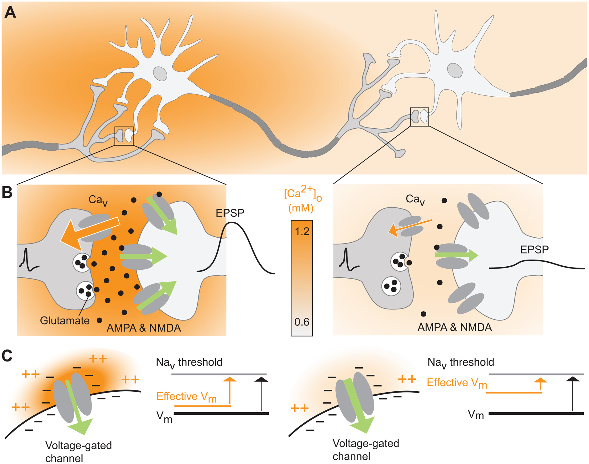
(A) Schematic diagram of a neural circuit characterized by an activity-dependent local decrease in [Ca2+]o. (B) Lowering [Ca2+]o can reduce the strength of synaptic signaling. As [Ca2+]o falls, the reduced driving force for Ca2+ influx through voltage-gated Ca2+ channels (Cav) leads to weakened presynaptic glutamate release, and, in extreme cases, action potential failure. Green arrow directions and widths reflect the direction and strength of the driving force for primarily Na+ influx; excitatory postsynaptic potential (EPSP) amplitudes are shown by schematic membrane potential traces. (C) The effective membrane potential across voltage-gated channels is partly governed by the high concentration of negatively charged residues on these channels’ interstitial domains. Under conditions of physiological [Ca2+]o (1.2 mM, left) the negatively-charged residues are largely neutralized by divalent cations (both Ca2+ and Mg2+). As a result, the local effective membrane potential is similar to the soma. Conversely, lowering the [Ca2+]o to, for example 0.6 mM as occurs during seizures, exposes the negative on the channels’ interstitial domains resulting in a local depolarizing the membrane and increasing neuronal activity through voltage-gated channels.