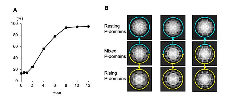
Fig 2. Transformation process of the dynamic rotation of P domain in MNoV-1 infectious particles.
(A) MNoV-1 particles having the rising P domain conformation in PBS(-)+EDTA (pH 8) were suspended in DMEM. The particles were then observed directly using cryo-EM. The particles showing the resting conformation in all P-domains were counted in the classes after 2D classification. The percentage of the MNoV-1 particles having the resting P domain conformation were plotted over time (n = ~200 particles in each point). (B) Representative 2D average images of MNoV-1 particles between 2 and 6 hours. Particles mixed with two conformations were observed (second row panels). The mixed P domain structure within a single particle suggests that even if the conversion of individual P domains may be rapid, it would take time to modify the overall conformation of the capsid in this aqueous condition, where the P domains are connected together like an entangled net.

