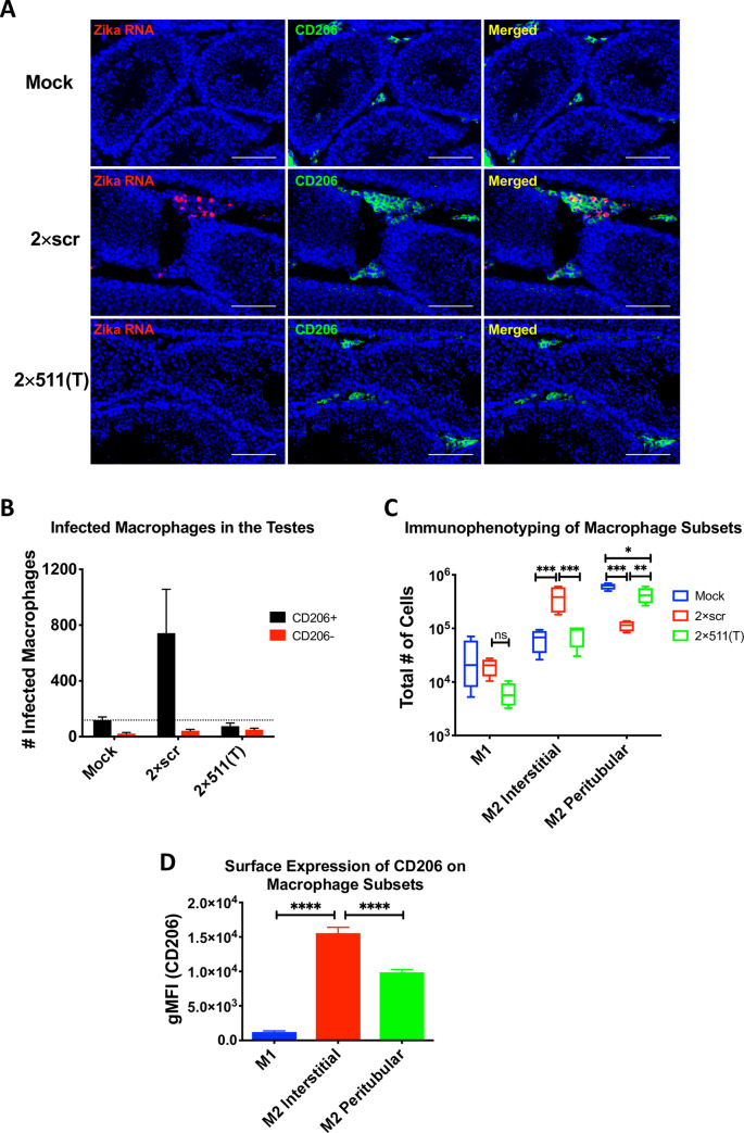
Fig 3. Targeting of ZIKV genome for mir-511-3p prevents infection of CD206 expressing macrophages in the testicular interstitium.
Adult AG129 male mice were mock-inoculated or infected IP with 106 pfu of 2×scr or 2×511(T) virus. For panel (A), mock (n = 1), 2×scr (n = 3) and 2×511(T) (n = 3); for panels (B-D), mock (n = 3), 2×scr (n = 3) and 2×511(T) (n = 4). Mice were sacrificed at 3 dpi and testes were harvested. (A) Testes sections stained for ZIKV RNA (red) by in situ hybridization and CD206 (green) by immunofluorescence co-staining. Scale bars represent 100μm. To identify the ZIKV-infected cells within the CD206-positive and CD206-negative populations, cells from testes at 3 dpi were dissociated into a single cell suspension, fixed, and stained for ZIKV using anti-E protein antibody, 4G2, to detect ZIKV and CD206 (B). Single cell suspensions from testes were stained with CD45, F4/80, CD11c, CD11b, MHC II and CD206 to differentiate various myeloid cell types (C). (D) Expression of CD206 was determined in each cell population identified in C. Statistical significance was determined for (C) and (D) by two way ANOVA and one way ANOVA for multiple comparisons. *, **, ***, **** indicate p<0.05, p<0.01, p≤0.001 and p≤0.0001, respectively.

