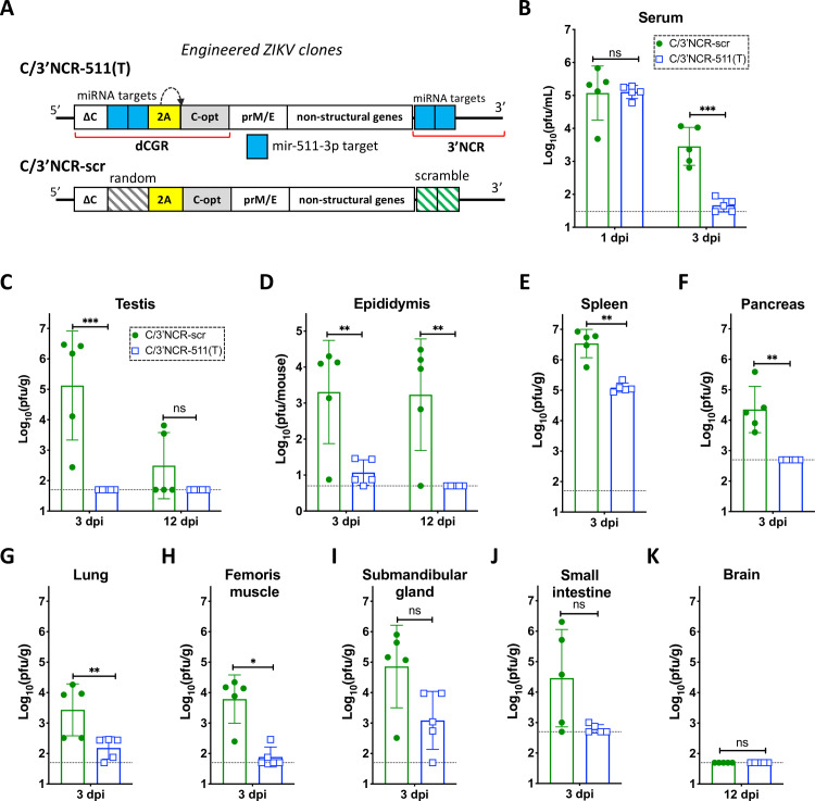
Fig 6. Insertion of multiple copies of mir-511-3p target sequences into distant regions of the ZIKV genome inhibits viral replication in peripheral mouse organs.
(A) Schematic representation of viruses used in the mouse studies. (B-K) Male AG129 mice (5 per group) were infected IP with 106 pfu of C/3’NCR-511(T) or C/3’NCR-scr virus and were bled at 1 dpi and sacrificed at 3 dpi, or were not bled and sacrificed at 12 dpi. (B) Mean virus titer ± SD in the serum at 1 and 3 dpi. (C-D) Mean virus titer ± SD in the testes (C) and epididymis (D) at 3 dpi and 12 dpi. (E-K) Mean virus titer ± SD in the spleen (E), pancreas (F), lung (G), femoris muscle (H), submandibular gland (I) and small intestine (J) at 3 dpi, and in the brain (K) at 12 dpi. The dashed lines indicate the limit of virus detection: in the serum [1.5 log10 pfu/mL]; in the testes, spleen, lung, femoris muscle, submandibular gland and brain [1.7 log10 pfu/g of tissue]; in the pancreas and small intestine [2.7 log10 pfu/g of tissue]; and in the epididymis [0.7 log10 pfu/mouse]. Differences between viral titers in the serum, testes and epididymis were compared using two-way ANOVA, and differences between viral titers in other organs were compared using two-tailed Mann-Whitney test. *** p<0.001; **** p< 0.0001; ns denotes p>0.05.

