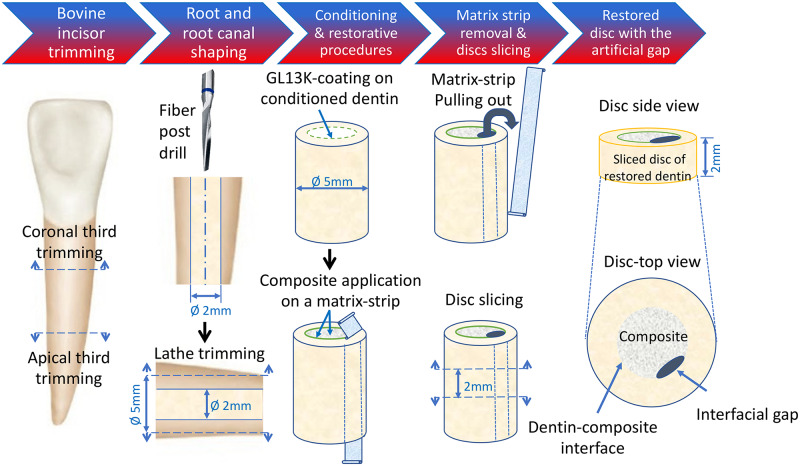
Fig 1. Schematics for the preparation of an ex vivo simulated dentin-composite failed interface used to assess the impact of AAMPs-treated interfaces on dental plaque biofilms.
A reproducible artificial gap was introduced along the dentin-composite interface of the restored discs using a method adapted from [25]. The radicular dentin tubes were total-etched and filled with dental resin composite (Filtek™ Z250 Universal Restorative 3M, St. Paul, MN, USA) without adhesive. Clear matrix strips (4" x 3/8" x 0.002") (Patterson® Mylar® Matrix Strips, Patterson Dental, Dental Supply, St. Paul, MN, USA) were introduced in the dentin cylinders, against the dentin wall, before composite application. After curing of the composite, the matrix strips were pulled out leaving a gap of ~ 400 μm in thickness along the interface. Then, the restored dentin cylinders were transversally sliced using a pre-calibrated template into 2-mm thick dentin-composite discs. This model was used to assess the biofilm succession along the interface with and without D-GL13K treatment using multiphoton deep bioimaging as detailed in Fig 5.

