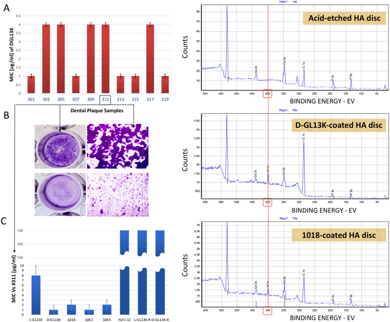
Fig 2. Antimicrobial effect of peptide candidates on dental plaque communities.
A) The minimum inhibitory concentrations (MICs) of D-GL13K against 10 different supragingival plaque samples collected from caries active individuals. Bars show average ± standard deviation (n = 3). B) 6-day-old highly adherent plaque biofilm of #311 sample (top panels). Bottom panels show #311 sample treated with 25μg/ml D-GL13K [22]. C) MICs of antimicrobial and non-antimicrobial control peptides against #311 plaque sample [23]. Bars show average ± standard deviation (n = 3). D) X-ray photoelectron spectroscopy (XPS) spectra for peptide coatings on HA discs. Top panel shows the surface atomic composition of acid-etched (32% H3PO4) control HA discs. Middle and bottom panels show the surface atomic composition of etched HA discs after coating with D-GL13K (an all D-amino acids peptide) and 1018 (an all L-amino acids peptide), respectively. The red dashed line shows the position of the nitrogen-N1s peak. The appearance of the nitrogen peak in peptide-coated HA discs is indicative of the presence of the peptides on these discs. HA: Hydroxyapatite.

