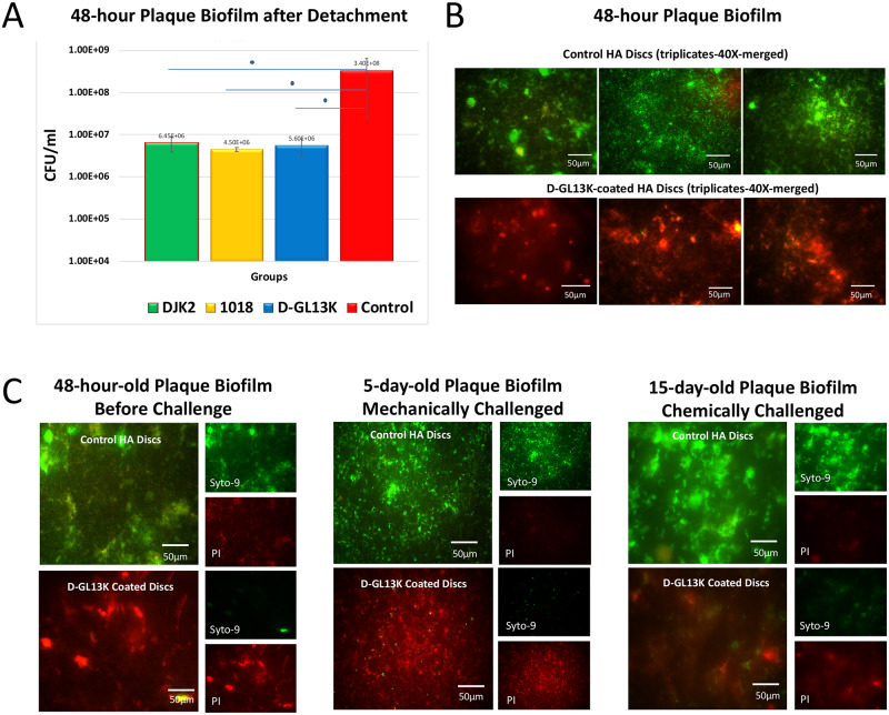
Fig 3. Antibiofilm effect of D-GL13K, DJK2, and 1018 coated HA discs on dental plaque biofilms.
A) Cell death quantification by counting colony forming units (CFU) for detached bacteria biofilms from HA discs coated with all AAMPs. Control group is 32% phosphoric acid-etched non-coated HA discs. N = 3. B) Merged live (green) and dead (red) bacteria viability assay images of 48-hour plaque biofilms grown on HA discs with (bottom image) and without (top image) D-GL13K coatings. C) Live and dead viability assay images of 48-hour (left), 5-day (middle), and 15-day-old (right) plaque biofilms grown on HA discs with (bottom images) and without (top images) D-GL13K coating. Middle panel shows regrown biofilms for 5 days after 45 min of ultrasonication (mechanically challenged D-GL13K coating). Right panel shows regrown biofilms for 15 days after 45 min immersion in 30% acetic acid (chemically challenged D-GL13K coating). Merged images are on the left side of each group and separated live (top)/dead (bottom) bacteria images are on the right side of each group.

