
Fig 5. Detection of PD-1 expression on CD4+ and CD8+ T-cells from peripheral blood by anti-canine PD-1 antibody JC053.
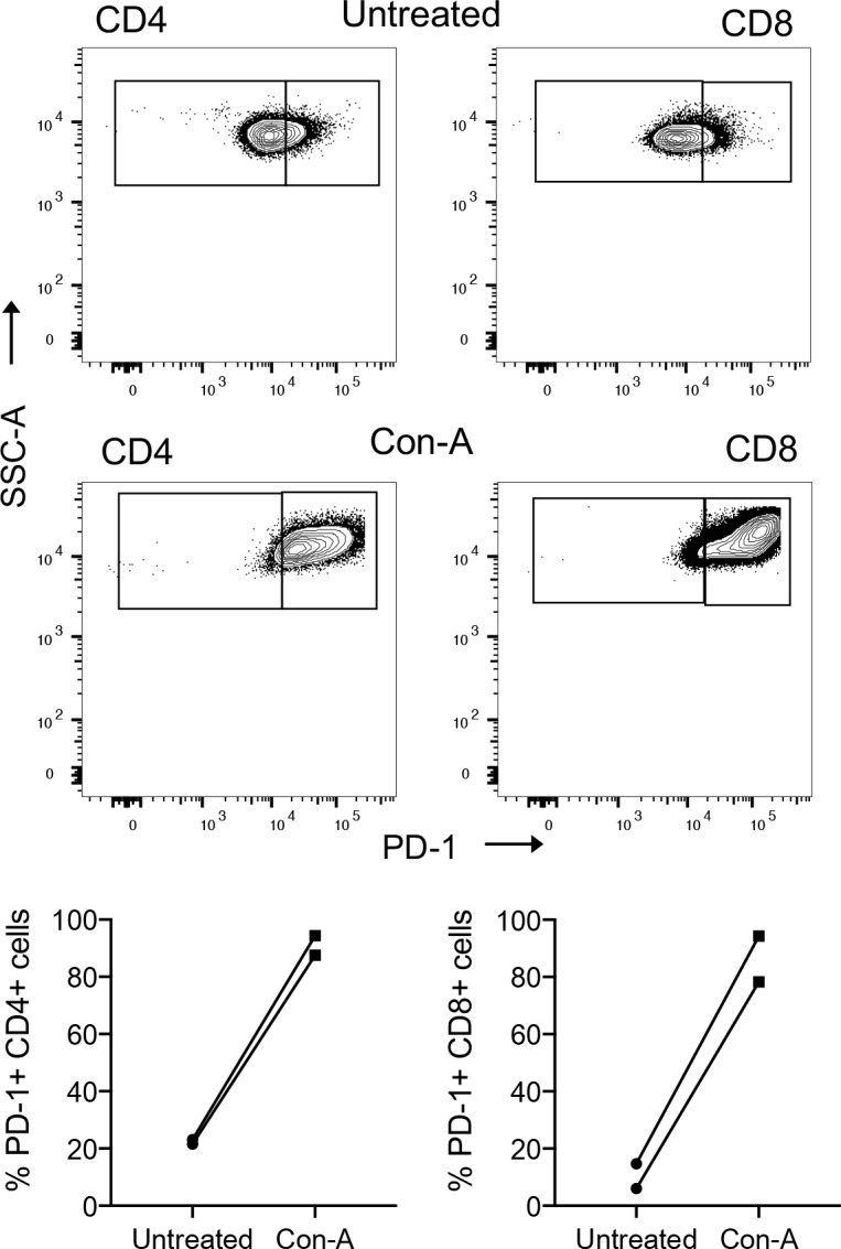
Representative staining of PBMC T cell subsets for PD-1 expression by flow cytometry using antibody JC053 from a healthy canine donor is shown before and after stimulation with ConA for 24 hours (A). Frequencies of PD-1 expressing CD4+ and CD8+ T-cells before and after stimulation of PBMCs from two healthy canine donors were evaluated by flow cytometry using antibody JC053.