
Fig. 1. Uninterrupted Recovery Experiment Results.
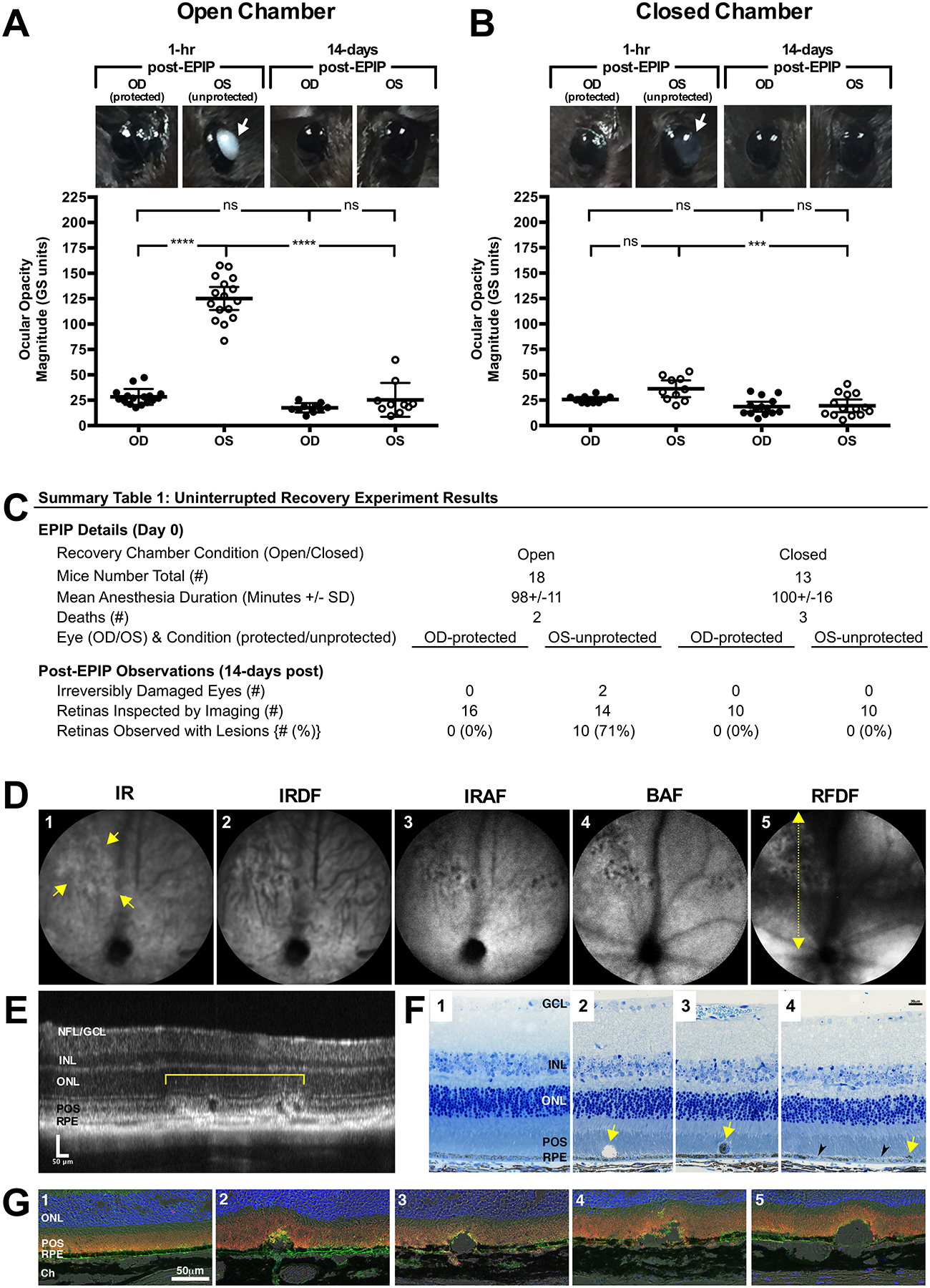
(A & B) C57BL/6J and Tulp1+/+ mouse eyes that receive no form of ocular protection developed lens media opacities that could be visualized by the naked eye. Opacities were worse in the exposed (OS) eyes of mice recovered in the open (A) vs. closed (B) chamber at 1-hr post-EPIP. For the most part, these opacities resolved by 14-days post-recovery with exception to two instances of irreversibly damaged eyes that resulted in microphthalmia. Eyes that were covered with protective ointment and eye shields did not develop any significant lens media opacities regardless of whether the recovery chamber was open or closed. Arrows indicate the eyes with visible opacities. Note the distinctive difference in the media opacity appearance between mice recovered under high-humidity conditions vs. a typical room environment with lower humidity levels. (C) Experimental details and a summary table of the observations made for fundus imaging of C57BL/6J and Tulp1+/+ mice at 14-days post-EPIP. (D) Representative SLO images from a Tulp1+/+ mouse with retinal lesions using IR, IRDF, IRAF, BAF, and RFDF imaging modes. Yellow arrows indicate the margins of a retinal lesion observed 14-days post EPIP recovery. RFDF (D; yellow dotted-line w/arrows) image indicates the approximate location of the SD-OCT B-scan shown (E) that was collected through an SLO detected lesion. Dark spots in BAF-SLO image are indicative of the cysts found subsequently by SD-OCT imaging (E) and histology (F2 & G2−5). (F) Photomicrographs of one regular (F1) and three abnormal (F2−4) thin section histology examples collected from one unaffected and three individually or affected mice, respectively. F2 shows an enlarged cyst-like structure above the RPE that is mostly devoid of material with exception to several pigment granules. F3 shows a detached, nucleated cell filled with pigment. F4 shows clustered pigment (yellow arrows) and hypopigmented (black arrowheads) regions of RPE. Immunohistomicrographs of one regular (G1) and four abnormal (G2−5) examples collected from one unaffected and four individual affected mice, respectively (Blue-TOPRO-3, Green-GLUT1, & Orange-Rhodopsin). (G2−5) Subretinal cyst-like structures and disruptions to the photoreceptor outer segments and interface with the RPE are readily visible. Perturbations as large as 50 μm can be seen displacing photoreceptor inner and outer segment lamina in the vitreal direction. OCT/Histology Abbreviations: Nerve Fiber Layer (NFL), Ganglion Cell Layer (GCL), Inner Nuclear Layer (INL), Outer Nuclear Layer (ONL), Photoreceptor Outer Segments (POS), Retinal Pigment Epithelium (RPE), and Choroid (Ch).