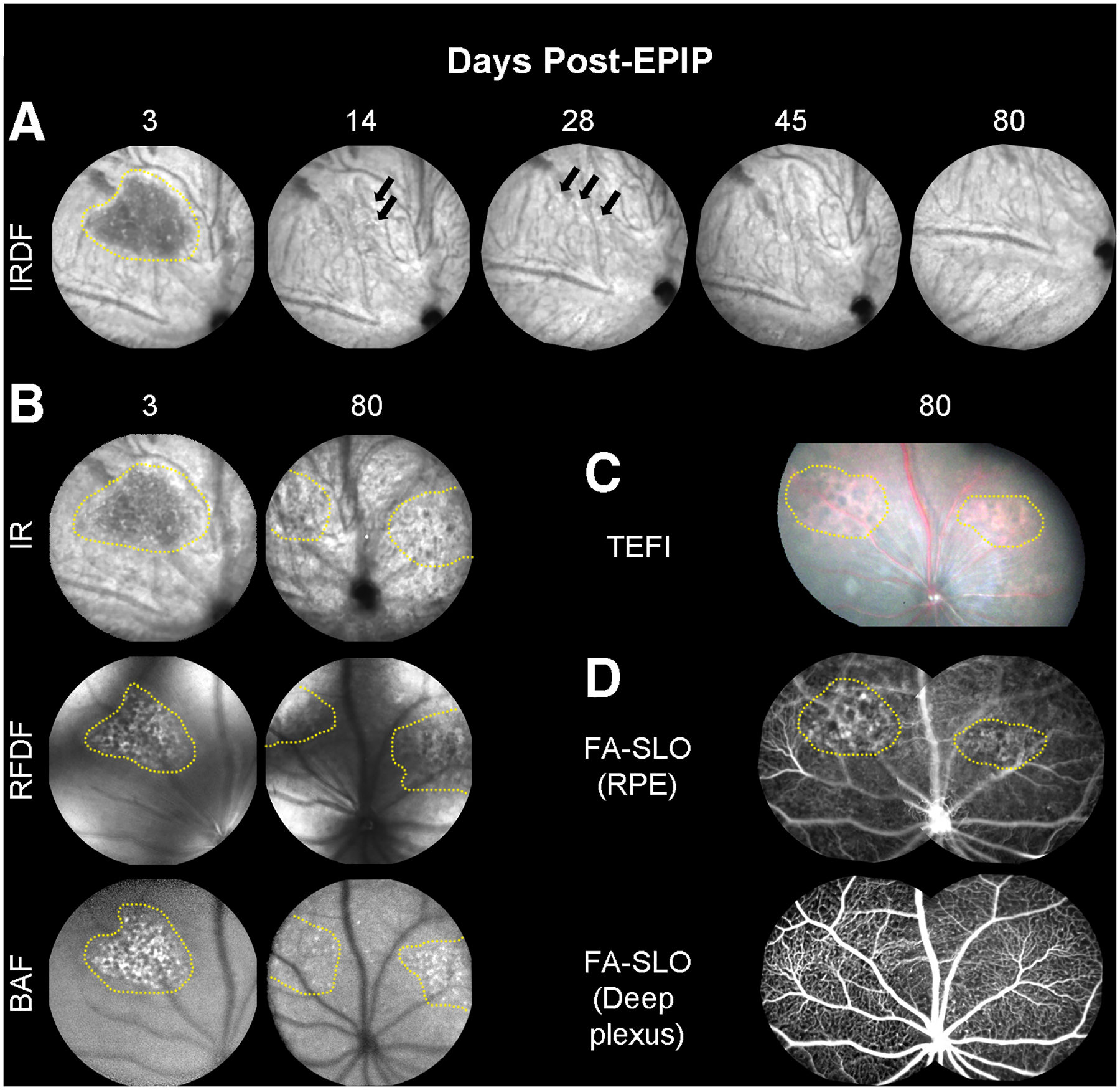
Fig. 4. Long-term follow up of retinal lesions.

(A) Representative IRDF-SLO images of lesions in a C57Bl/6J mouse at 3-days post-EPIP, which are easily discernable from the standard RPE/choroidal background (n = 3)… The lesion is presented as a dark region that has resolved 14-days post-EPIP. At 14 and 28-days, subtle indicators (hyper- and hypo-reflective spots) persist that suggest some evidence of the lesion remains. By 45 and 80-days, the hyper- and hypo-reflective perturbations have resolved, and the region appears similar to the surrounding background. (B) Additional examples of the same lesion shown by IRDF (Fig. 4A) at 3-days post are also readily visible by three of the other SLO imaging modes (IR, RFDF, and BAF). This comparison helps to identify and isolate the region impacted by the lesion, which appears to be the outer retina and RPE. As a result of this observation, it is suggestive that the lesion remaining at 80 days post-EPIP is also altered RPE and this is further supported by the TEFI and FA-SLO images that follow. (C) Color fundus images showing a different spectral reflectance profile for the two visible lesions versus the surrounding background. (D) Sodium Fluorescein Angiography (FA-SLO) revealing leakage or abnormal uptake of the fluorophore at the RPE level whereas no evidence of leakage can be observed in the deep plexus of the retinal vasculature. The enhanced visualization of red reflectance and green fluorescence for the TEFI and FA-SLO images, respectively, could alternatively be due to hypo-pigmentation of the RPE as previously indicated in Fig. 1F4. These observations suggest that the damage remaining at this late stage is isolated to the RPE.
