
Fig. 5. Anterior Segment SDOCT Imaging Examples and Quantified Results.
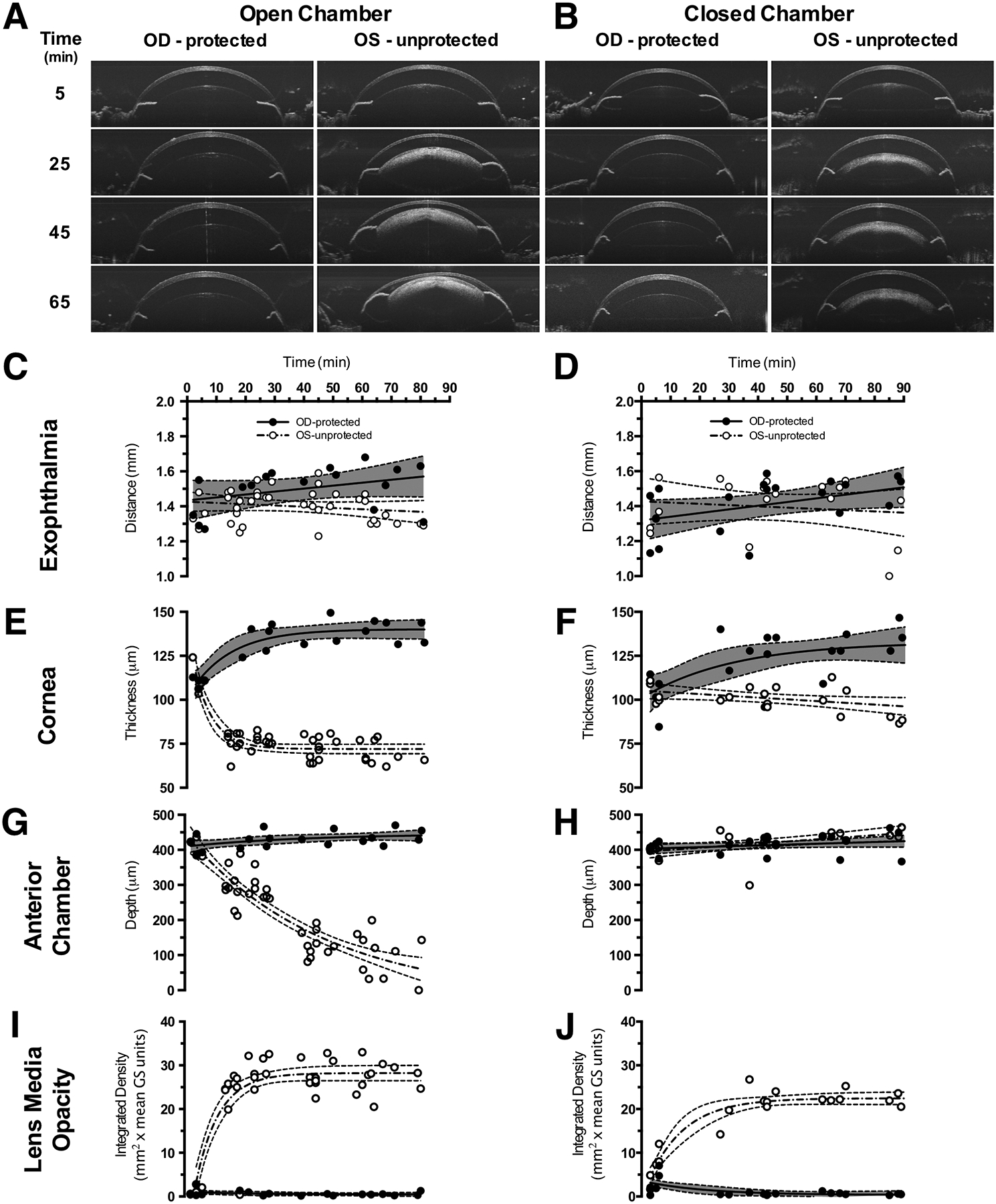
Representative SDOCT images from the anterior segment of Tulp1+/+ mice (n = 8) recovered in the open (A) vs. closed (B) chambers. Eyes of mice that received ocular protection (OD-protected) exhibited no substantial adverse changes compared to eyes that were left unprotected (OS-protected). However, mice with unprotected eyes (OS-unprotected) and recovered in the closed chamber (B) only developed lens media opacities in contrast to mice with unprotected eyes (OS-unprotected) and recovered in the open chamber that (A) developed visibly apparent corneal thinning, lens media opacities and reduction in anterior segment depth. Quantitative results for exophthalmia (CD), corneal thickness (E-F), anterior chamber depth (GH) and lens media opacity (I-J) for the two ocular status and two recovery and conditions.