
Fig. 6. Rate of Change Comparisons.
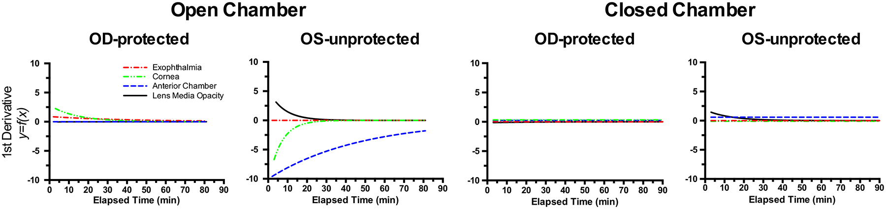
First derivatives taken of the fitted curve trends shown in Fig. 5 reveal the rate of change for exophthalmia, corneal thinning, anterior chamber depth collapse, and ocular lens media opacity development. These curves show the magnitude, direction, and rate of change associated with these metrics measured in vivo via anterior segment SD-OCT imaging. Note that the most prevalent changes occur in the unprotected left eyes (OS-unprotected) of mice that are recovered in the open chamber. Furthermore, anterior chamber depth is observed having the largest magnitude and most sustained rate of change during the 80 min of monitoring.