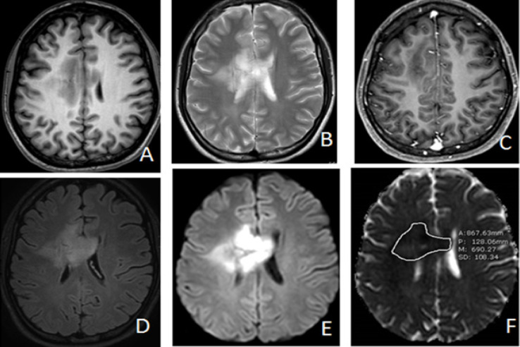
Figure 3.
Anaplastic Astrocytoma (WHO Grade III) in 19 Year-Old-Woman. Axial T1W (A), T2W(B), Post contrast T1WFS(C), FLAIR (D), DWI(E) and ADC map (F) images representing case that showed marked hyperintense solid part of the tumor with visual scale 5 on DWI (E) and ADC value measurement = 690.27 x 10⁻⁶ mm2/s on ADC map(F) which is below the cutoff value

