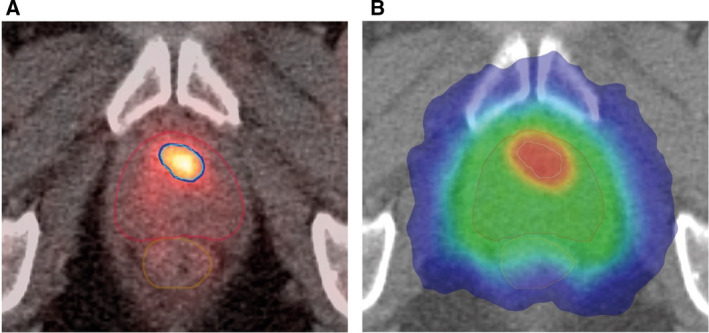
Fig. 4.

PSMA‐PET‐based focal boosting in prostate cancer. (A) Axial PSMA‐PET‐CT slice showing the contours of the prostate (red), GTV (cyan), rectum (brown), and the 50 Gy isodose (5 fractions of 10 Gy; marine blue). (B) Corresponding CT slice with color wash isodose curve showing conformal dose shaping to the prostate (clinical tumor volume) treated to 35 Gy in five fractions of 7 Gy and intraprostatic tumor (GTV) with sparing of the rectum and urethra. The intraprostatic lesion (cT1c, Gleason 3 + 4 = 7, iPSA = 16.6 ng·mL−1) is located in the left transition zone. The patient participated into the multicenter prospective phase II hypo‐FLAME study (NCT02853110, ClinicalTrials.gov).
