FIG. 2.
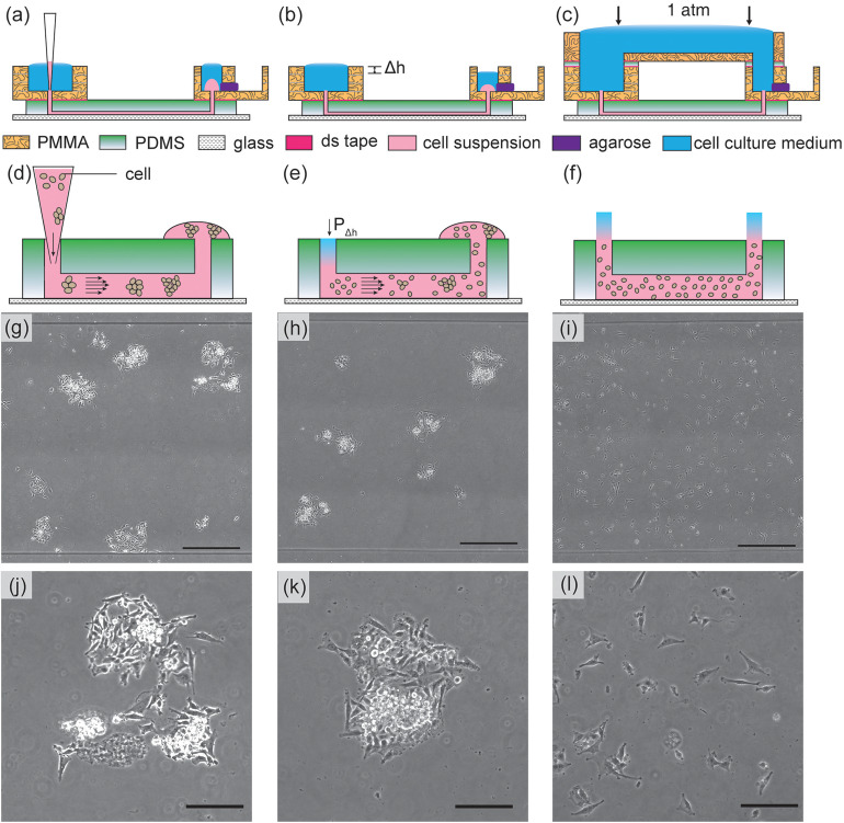
The results of U-251MG cell seeding inside microchannels by various methods. [Left column: (a), (d), (g), and (j)]. In the tip loading method, cells are introduced by using gravitational flow with micropipette tips. The cells can flocculate inside the tips and in microchannels as illustrated in (d). The microscopy image of seeded cells is shown in (g) and magnified in (j); [Middle column: (b), (e), (h), and (k)] in the tip injection method, cells are injected into the channels and tips are removed. The small hydrostatic pressure differences between the inlet/outlet (shown as Δh) will contribute to hydrodynamic flow and disturb the cell distribution, causing non-uniform cell distribution and aggregates as shown in (e). The microscopy image of seeded cells is shown in (h) and magnified in (k); [Right column: (c), (f), (i), and (l)] in our pressure-balanced submerged cell seeding method, the hydrostatic pressure difference is eliminated. The injected cells remain uniform throughout the channel as shown in (f). The microscopy image of seeded cells is shown in (i) and magnified in (l). The uniform and sparse cell seeding method is suitable for different applications such as single cell tracking, ensembled cell studies, and cell assembly. The scale bars in [(g), (h), and (i)] represent 500 μm. The scale bars in [(j), (k), and (l)] represent 200 μm.

