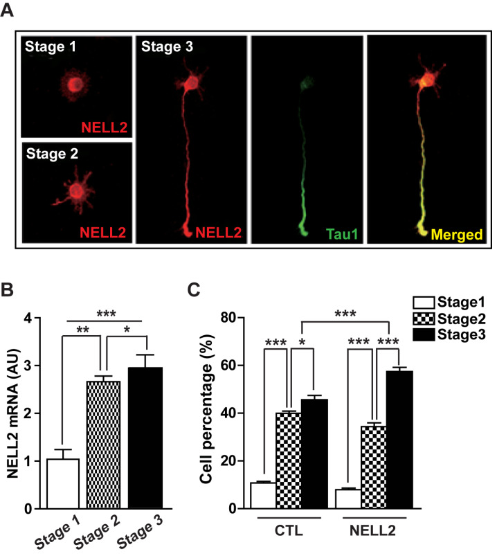
Fig. 1. NELL2 function in the differentiation of cultured hippocampal neurons.
(A) Expression of NELL2 in cultured hippocampal neurons during stages 1 to 3. Hippocampal neurons were stained with anti-NELL2 and anti-Tau1 antibodies. Scale bar = 20 µm. (B) Change of NELL2 mRNA level in cultured hippocampal neurons during developmental stages 1 to 3. (C) Effect of NELL2 on the development of cultured hippocampal neurons. Neurons were transfected with control (pDS-GFP-XB, CTL) or NELL2-overexpresion vector (pDS-NELL2-GFP, NELL2). The progression of developmental stage was observed in the transfected cells that were identified with fluorescence. All data are presented as mean ± SEM. n = 60 (CTL) and 62 (NELL2) cells. *P < 0.05; **P < 0.01; ***P < 0.001. AU, arbitrary units. P values for unpaired comparisons were analyzed by two-tailed Student’s t-test. Two-way repeated-measures ANOVA was performed to detect significant interaction between groups.

