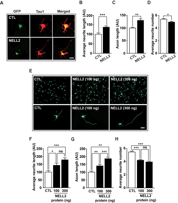
Fig. 2. Effect of NELL2 on neuronal polarization.
(A) Representative microphotographs of immunocytochemistry. Hippocampal neurons were cultured and transfected with pDS-GFP-XB (CTL) or pDS-NELL2-GFP (NELL2) vectors. Neurons were fixed at 2 days after transfection and stained with anti-Tau1 antibody. (B-D) Hippocampal primary cells transfected with CTL or NELL2 vectors were analyzed to determine the average neurite length (B), axon length (C), and average neurite number (D). All data are presented as mean ± SEM. n = 41 (CTL) and 48 (NELL2) cells. (E-H) Hippocampal primary cells were treated with human NELL2 proteins and their neuronal polarization was analyzed after staining with anti-Tau1 antibody: representative microphotographs (E), neurite length (F), axon length (G), and average neurite number number (H). Scale bars = 20 µm (A and E). n = 35 (CTL), 48 (NELL2, 100 ng), and 40 (NELL2, 300 ng) cells. *P < 0.05; **P < 0.01; ***P < 0.001; ns, no significance. AU, arbitrary units. P values for unpaired comparisons were analyzed by two-tailed Student’s t-test.

