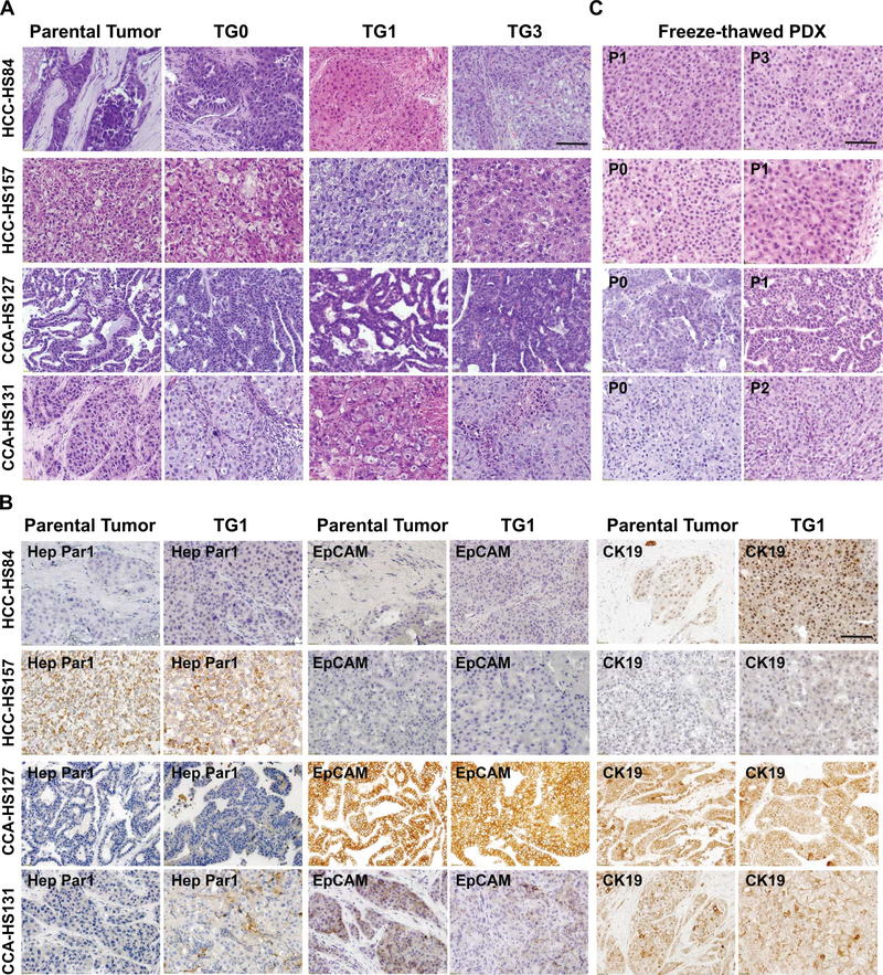
Figure 4. PDXs maintain the features of parental tumor histology.
A. H&E staining of parental tumors and passaged PDX lines. Scale bar = 100μm.
B. IHC staining of parental tumor samples and PDXs with anti-Hep Par1, anti-EpCAM, and anti-CK19 antibodies. Scale bar = 100μm.
C. H&E staining showed that thawed PDXs have the similar histology as the primary PDX and patient tumor. Scale bar = 100μm.
H&E = Hematoxylin and eosin; TG = Tumor Graft

