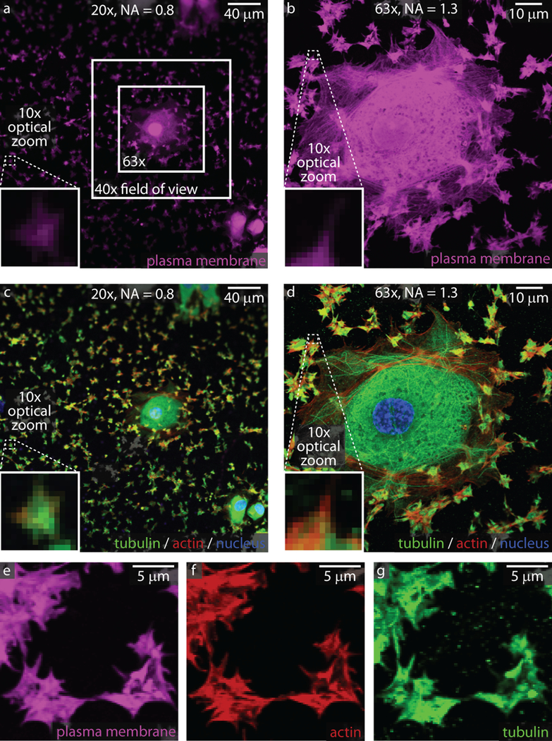
Figure 1: Imaging considerations for single platelets and aggregates.
Platelets are much smaller than most cells in the body, as can be seen next to a spread endothelial cell adhered to collagen and stained with a plasma membrane dye (a, b) or with actin/tubulin staining (c, d). At 20x, the general platelet morphology can be discerned. At higher magnifications and resolutions (63x), subcellular structures such as filipodia are visible with plasma dyes (b and e) or actin/tubulin staining (d, f, and g). Nuclei for the endothelial cell are shown in blue in panels c and d.

