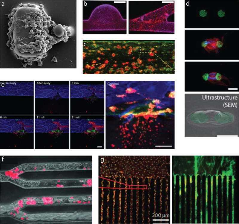
Figure 3: Next generation in vitro platelet imaging systems incorporate flow to measure dynamic functions of platelets in vitro.
(a) The combined aggregation and force potential of flowing platelets is measured by measuring the deformation of the smaller post [54]. (b) Various geometries covered by endothelial cells enable studies of how geometry, flow, and the presence of an endothelium influence platelet aggregation[60]. (c) More portable devices utilizing fixed endothelial cells can also measure platelet function[62]. (d) Single platelet contraction forces can be measured in a high-throughput manner, using large numbers of these arrays under flow conditions in microfluidic devices[8]. (e) Integrating valves into microfluidic channels with endothelial cells can even lead to a system that can be injured and “bleed”, which is useful for modeling hemostasis[61]. (f) Multi-channel microfluidic devices reveal that platelets can interact with endothelial cells in numerous geometries[58] and can be used to (g) image multiple shear conditions simultaneously[36].

