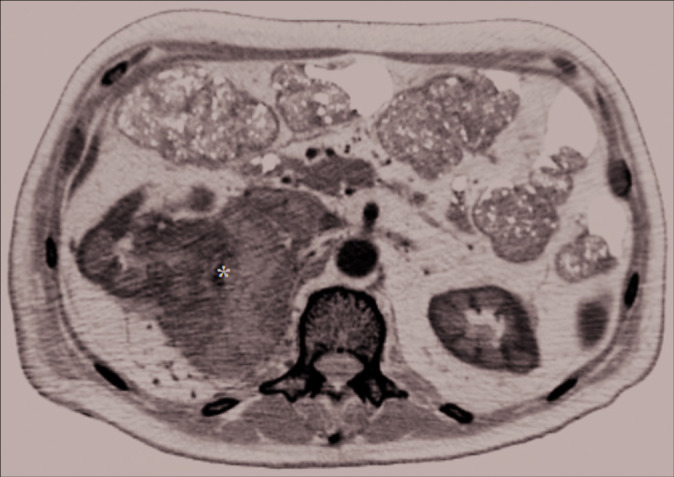
Figure 2:

Axial abdominal contrast-enhanced computed tomography scan image showing a voluminous mass (about 85 mm) (black asterisk) involving the upper polar region and the middle third of the right kidney, the ipsilateral adrenal gland, and extends posteriorly to infiltrate the ipsilateral psoas muscle. This lesion, which presents an inhomogeneous hypodense aspect with hypervascular foci in this context, is associated with collateral circles in the peri- and pararenal space, with the infiltration of the upper right calyxes. A neoplastic thrombosis of the renal vein and inferior vena cava in the subhepatic tract is also present and may explain hematogenous spread through Batson’s venous plexus.
