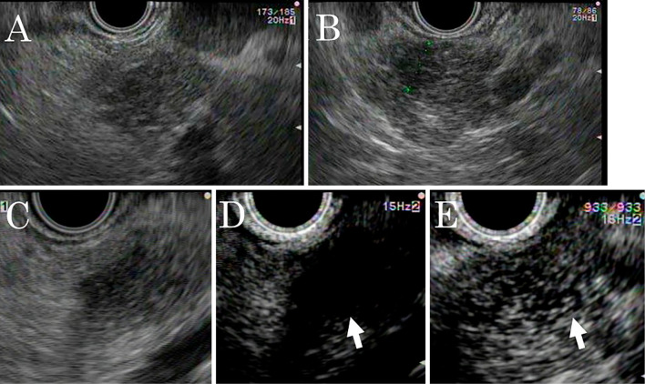
Figure 5.
Endoscopic ultrasonography (EUS) findings in a 52-year-old man with multiple lesions in the body and tail of the pancreas. A, B: Conventional EUS reveals several isodense, ill-defined, poorly-demarcated, hypoechoic lesions in the pancreatic body and tail. A: Pancreatic body, B: Pancreatic tail. C, D, E: Enhanced EUS using an ultrasound contrast agent. C: Pre-injection. A low-echoic lesion. D: Twenty seconds post-injection. The lesions are less enhanced than the pancreatic parenchyma (arrow). E: After 60 s, the lesions became isoenhanced and ill-demarcated from the pancreatic parenchyma (arrow).

