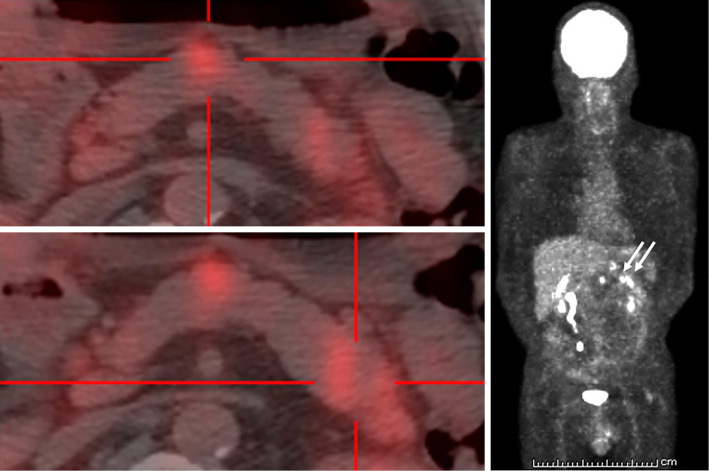
Figure 7.
Fluorine-18-deoxyglucose (FDG)-positron emission tomography/CT in a 52-year-old man with multiple lesions in the body and tail of the pancreas. Several lesions in the body and tail of the pancreas are visible (arrow), but no lesions in other organs are seen. A: The accumulation in the pancreatic body. B: The accumulation in the pancreatic tail. C: Coronal section

