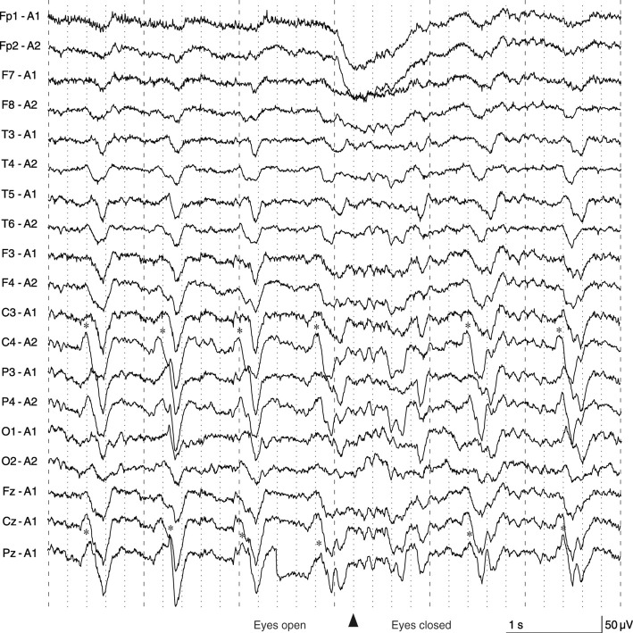
Figure 2.
Electroencephalogram findings. PSWCs (asterisks) were observed in a region extending from the central area to the parietal area. PSWCs in the right hemisphere preceded those on the left side by approximately 0.1 seconds, which indicated that the PSWCs originated from the right hemisphere and spread to the left hemisphere. Posterior dominant rhythms were slow (8 Hz) on both sides and suppressed when the eyes were open. The patient exhibited left-side EPC during the test, but we did not perform simultaneous recording of electromyography or jerk-locked back averaging. EPC: epilepsia partialis continua, PSWC: periodic sharp wave complex

