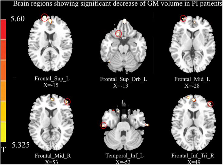
FIGURE 1.
Compared with healthy controls (p < 0.05, FEW corrected), the volume of gray matter (GM) in the left dorsolateral prefrontal cortex (DLPFC), left orbital prefrontal cortex (OFC), bilateral middle frontal gyrus (MFC), left inferior temporal gyrus, and right inferior frontal gyrus (IFG) was decreased in primary insomnia (PI) patients. No increased GM volume was observed in the brain regions of PI patients in this study.

