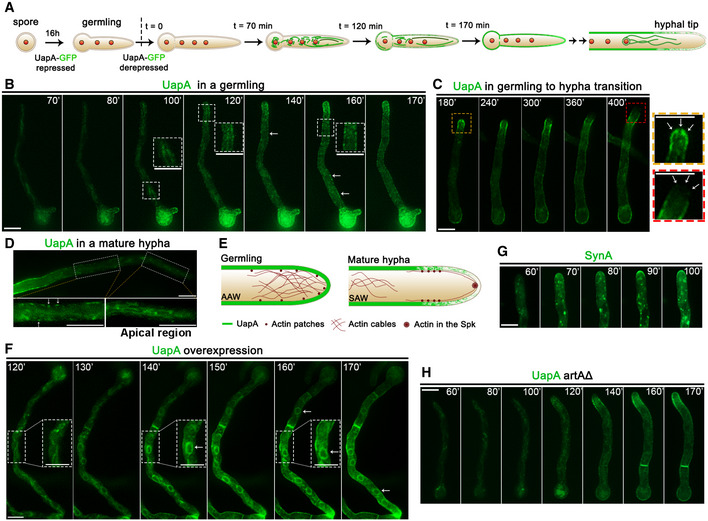
Figure 1. Subcellular localization of neosynthesized UapA.

-
ACartoon depicting the strategy for following the trafficking of neosynthesized UapA. The subcellular localization of UapA is shown in green. Red circles indicate positioning of nuclei. (for more details, see main text).
-
BIn vivo epifluorescence microscopy following de novo expressed UapA‐GFP in a single growing germling at 70, 80, 100, 120, 140, 160 and 170 min after derepression of transcription via its native uapA promoter (for details, see text and Materials and Methods). Notice that in the growing germling tip UapA localizes in a membranous mesh and some cytosolic puncta, whereas in more tip‐distal parts of the germlings, UapA is in PM‐localized puncta (see white arrows), which progressively become more abundant so that at more posterior areas the PM is homogeneously labelled.
-
CIn vivo epifluorescence microscopy following de novo expressed UapA‐GFP in a germling maturing to young hypha at 180, 240, 300, 360 and 400 min after derepression of transcription via its native uapA promoter. During this developmental transition, UapA apical localization is gradually diminished. As the cell tip acquires a faster growth rate, UapA is completely retracted from the hyphal apex (see zoom‐in panels on the right and white arrows).
-
DIn a mature hyphal cell, UapA is no longer found in the PM of the extreme apical compartment, but in a cytoplasmic membrane network resembling the ER. As the distance from the apex increases, UapA progressively populates the PM as cortical puncta (see white arrows).
-
ECartoon depicting the rearrangement of actin and UapA at the tip of A. nidulans during germling to hyphal maturation. In germlings, dense arrays of actin cables are present in the apex, known as apical actin array (AAA). Actin patches are concentrated, but not restricted, to the apex. In mature hyphae, a core of actin appears in the position of the Spitzenkorper and actin patches are restricted to the subapical endocytic collar (≈2 μm behind the apex). Actin cables are retracted from the apex and shifted to a more distal region of the hyphal tip, the subapical actin web (SAW). In the apex, actin cables are now found associated mostly as sparse arrays across the cell cortex. The localization of UapA during hyphal maturation seems to follow the pattern of actin cables 26, 27.
-
FIn vivo epifluorescence microscopy following neosynthesized alcA p‐UapA‐GFP in a single hypha at 120, 130, 140, 150, 160 and 170 min, under overexpressing (derepression/ethanol‐induction) conditions, as described in Materials and Methods. Notice that UapA labels the perinuclear ER rings (white arrows).
-
GIn vivo epifluorescence microscopy following de novo expressed alcA p‐GFP‐SynA in a single young hyphal cell, at 60, 70, 80, 90 and 100 min after transcriptional derepression (for strain details, see Materials and Methods). SynA is a standard polar membrane cargo that traffics to the hyphal tip via Golgi‐dependent secretion. Notice the very distinct GFP fluorescent signals obtained using UapA (non‐polar membrane network and PM) versus SynA (Golgi‐like puncta and polar depositioning at the tip; see also alter).
-
HSimilar experiment as in (B) performed in an isogenic strain where ArtA, the arrestin required for UapA endocytosis, is genetically depleted.
