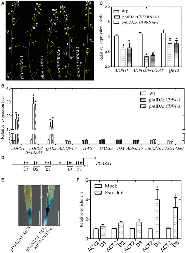
Figure 8. Altered floral organ abscission process in the CDF4 transgenic plants.

-
A–C(A) Observation of inflorescences of 7‐week‐old wild‐type (WT) Arabidopsis and the CDF4 transgenic plants. The asterisk represents the flower organ attached along the inflorescence. Scale bar indicates 1.5 cm. Selected abscission‐related gene expression levels in (B) WT and proIDA::CDF4 lines or (C) WT and proIDA::CDF4RNAi lines by using qPCR analysis. The expression of these selected genes in the wild‐type plant is given as 1. The relative expression level represents only the level of expression of the gene relative to the wild type. ACTIN2 was used as the internal control. Three independent experiments were conducted. Values are given as mean ± SD, n = 3. *P < 0.05 by Student's t‐test.
-
DSchematic diagram indicating the locations of the putative CDF4‐binding motif clusters (D1–D5) in the approximately 1.8‐kb promoter of PGAZAT.
-
EObservation of GUS staining activity in the proPGAZAT::GUS and proPGAZAT::GUS & proIDA::CDF4. Scale bar indicates 0.5 cm.
-
FChIP‐qPCR analysis of the relative binding of CDF4 to the promoter regions of PGAZAT. An anti‐HA monoclonal antibody was used for DNA immunoprecipitation from 6‐week‐old pER8::CDF4‐HA transgenic plants after estradiol induction. Black columns indicate the enrichment fold changes normalized to ACT2. Three independent experiments were conducted. Values are given as mean ± SD, n = 3. *P < 0.05 by Student's t‐test.
