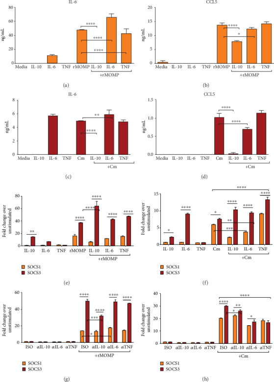
Figure 5.

The effect of exogenous and endogenously produced IL-6 and TNF on the expression of inflammatory mediators and SOCS in chlamydial-stimulated macrophages. Macrophages (106/mL) were stimulated with rMOMP (10 μg/mL) or Cm (MOI of 2) in the presence and absence of IL-10, IL-6, and TNF (each at 10 ng/mL) for 24 h. RNA and cell-free supernatants were collected to quantify IL-6 and CCL5 (A-D) along with SOCS1 and SOCS3 mRNA gene transcripts (E-F), respectively, by TaqMan qRT-PCR and specific ELISAs. Macrophages were pre-incubated with neutralizing Abs to IL-10, IL-6, and TNF (each at 25 μg/mL) for 30 min before adding rMOMP or Cm for an additional 24 h. Normal rat IgG1 Ab served as the isotype control (ISO). RNA was collected to quantify SOCS1 and SOCS3 mRNA gene transcripts (G-H). An asterisk indicates a significant difference (P <0.05), and P values were calculated as described in Figure 1. Each bar represents the mean ± SD of samples run in triplicates. Each experiment was repeated at least 3 times.
