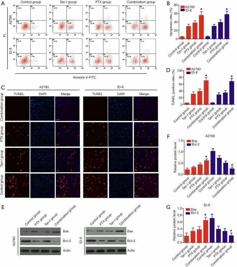
Figure 2.
Tan-I combined with Paclitaxel promote apoptosis of cancer cell. (A) Apoptosis assay of A2780 and ID-8 cells by flow cytometer in the control group, Tan-I group, PTX group, and combination group. A2780 and ID-8 cells were treated with Tan-I (4.8 μg/mL), Paclitaxel (0.1 μg/mL), or Tan-I combined with Paclitaxel for 24 hours. (B) The apoptosis-positive cells of A2780 and ID-8 cells in A. Data are shown as mean ± SD. *, P<0.05 versus which Tan-I group or Paclitaxel group. (C) Apoptosis assay of A2780 and ID-8 cells by TUNEL staining in control group, Tan-I group, PTX group, and combination group (magnification 100×). (D) The apoptosis-positive cells of A2780 and ID-8 cells in (C). Data are shown as mean ± SD. *, P<0.05 versus which Tan-I group or Paclitaxel group. (E) Bcl-2 and Bax expression level in A2780 and ID-8 cells by western blot in the control group, Tan-I group, PTX group, and combination group. (F) and (G) Relative fold change in the expression of Bcl-2 and Bax in comparison with Actin, and the results were analyzed by densitometric using image J software. Data are presented as the mean ± SD of three independent experiments. *, P<0.05 versus which Tan-I group or Paclitaxel group.

