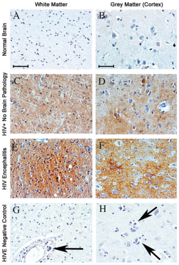
Fig. 1.

Immunohistochemical analyses of PINCH expression in HIV patients. All images are from frontal lobe and immunolabeled with anti-PINCH antibody (brown) and counterstained with hematoxylin. A,B: Expression of PINCH in the white and grey matter, respectively, of normal HIV-seronegative human brain samples is undetectable. C,D: In HIV-positive patients with no neurological complications, PINCH expression is increased with a patchy appearance in both the cortex and the subcotical white matter, respectively. E,F: In cases of HIVE, PINCH immunoreactivity is robust and widespread. G,H: In an HIVE case showing characteristic perivascular cuffing by mononuclear cells and a multinucleated giant cell (G, arrow) and cortical microglial nodule (H, arrows), primary anti-PINCH antibody was omitted to demonstrate antibody specificity. Scale bars = 10 μm.
