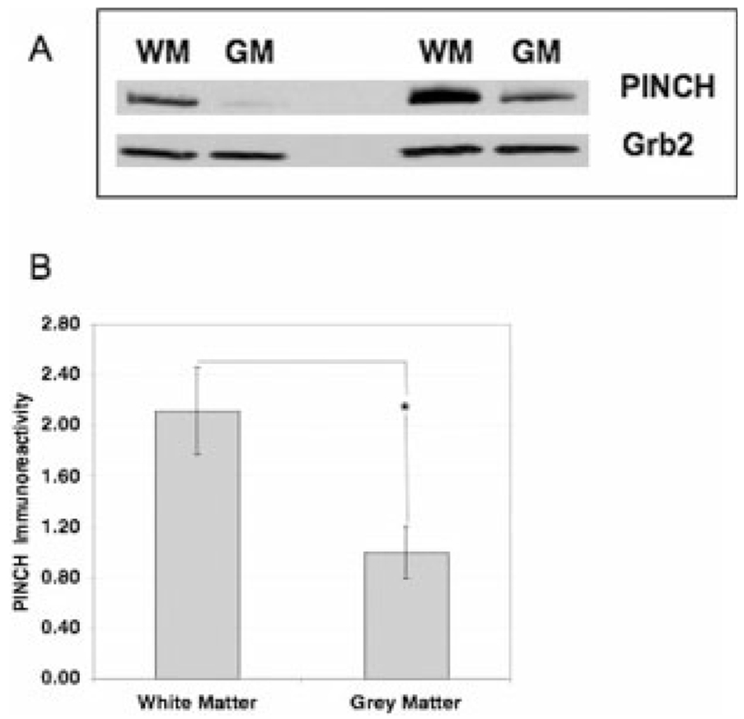
Fig. 4.

Western analyses of PINCH protein expression in white matter vs. grey matter. A: Western blot of frontal cortex homogenate from two representative HIVE cases enriched for either white matter (WM) or grey matter (GM) showing an immunoreactive band corresponding to the PINCH dimer at approximately 71 kDa. Grb2 was used as a loading control. B: Analyses of PINCH immunoreactivity in the white matter is significantly greater (*P < 0.001) than in the grey matter in HIVE cases (n = 14).
