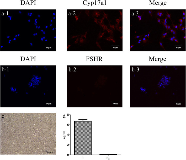
Fig. 2.
Immunostaining with Cyp17a1 and FSHR to characterize TICs (× 200). a1–a3 Cyp17a1 expression in TICs-blue fluorescence with DAPI nuclear staining and red fluorescence with Dylight 549 staining. b1–b3 FSHR expression in TICs-blue fluorescence with DAPI staining and red fluorescence with Dylight 549 staining (scale bar = 100 μm). c Microscopic morphology of TICs. d Levels of T and E2 in culture medium of TICs. Data are expressed as the means ± SD. FSHR, follicle-stimulating hormone receptor; TICs, theca-interstitial cells; DAPI 4, 6-diamino-2-phenyl indole; T, testosterone; E2, estradiol

