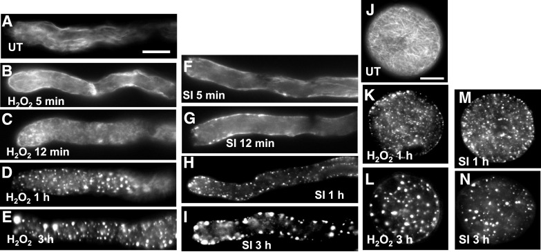
Figure 5.
F-actin alterations in pollen are induced by ROS in P. rhoeas pollen tubes. F-actin was visualized with rhodamine-phalloidin using fluorescence microscopy. A, F-actin organization in a representative untreated pollen tube. B to E, H2O2-treated pollen tubes after 5min (B), 12 min (C), 1 h (D), and 3 h (E) of treatment. Alterations were observed as early as 5 to 12 min after treatment. At 1 and 3h large punctate foci of actin were formed. F to I, Pollen tubes at 5 min (F), 12 min (G), 1 h (H), and 3 h (I) after SI induction showed similar alterations to F-actin. J to N, Pollen grains showed similar alterations. J, Untreated pollen grain with F-actin filament bundles; K and L, H2O2-treated pollen grains. M and N, SI-induced pollen grains. Scale bars = 10 μm. SI, SI induction; UT, untreated.

