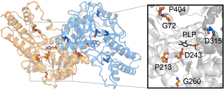
Figure 5.
Dimeric model of RTY. Locations of mutated residues and a detailed view of the PLP binding pocket in the RTY protein model are displayed. The mutations are labeled and shown in sphere-and-stick format on a cartoon-format background of the protein structure. One monomer is colored orange while the other is colored blue. PLP ligand, shown in stick-and-ball format, is included for clarity. The inset provides a detailed view of the substrate binding pocket and the location of the mutations that line the entrance and bottom of the PLP binding pocket. The surface is colored gray, while the mutations are colored orange and blue, and shown in sphere-and-stick format. The dimeric RTY model was generated by employing the MODELER software.

