Figure 2.
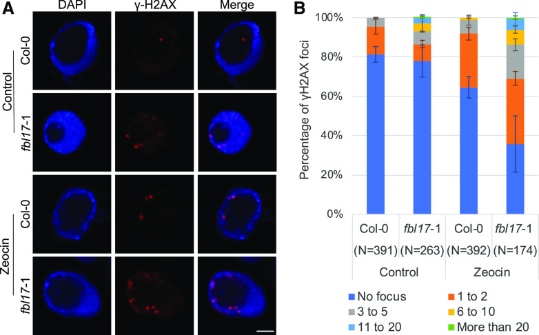
Increased accumulation of γH2AX foci in fbl17. A, Representative images of Col-0 and fbl17-1 after immunostaining of γH2AX foci (red) in root-tip nuclei from seedlings under control conditions or treated for 16 h with 5 μm zeocin. Nuclei were counterstained with 4′,6-diamino-phenylindole (blue). Scale bar = 2 μm. B, Quantification of γH2AX foci in nuclei from Col-0 and fbl17-1 seedlings under control conditions or treated with zeocin. Between 79 and 233 nuclei per line per replicate were analyzed and categorized into six types: no focus, 1 to 2, 3 to 5, 6 to 10, 11 to 20, or >20 γH2AX foci/nucleus, respectively. Two independent replicates were performed. Error bars indicate the sd.

