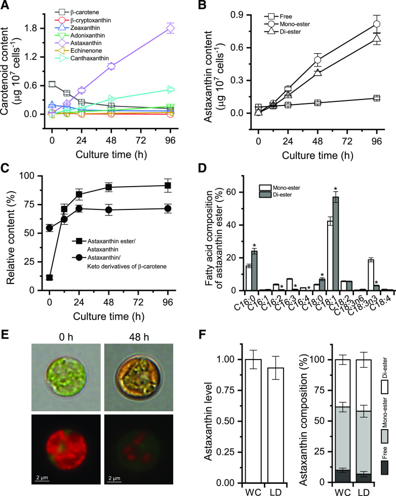
Figure 4.
Properties of astaxanthin synthesis and accumulation in C. zofingiensis. A, Time-course changes in the contents of β-carotene and its derivatives. B, Time courses of astaxanthin content in the form of free, monoester, and diester. C, Relative contents of astaxanthin ester to astaxanthin and of astaxanthin to keto derivatives of β-carotene. D, Fatty acid composition of astaxanthin monoester and diester at 48 h of ND. Asterisks indicate significant differences (Student’s t test, *P < 0.05) between monoester and diester. E, Microscopic views of C. zofingiensis cells at 0 and 48 h of ND. Top row, Bright field; bottom row, fluorescent field. Red indicates chlorophyll autofluorescence, whereas green indicates LDs stained with BODIPY. F, Comparison of astaxanthin levels and composition from whole-cell (WC) and LD fractions of C. zofingiensis cells after 48 h of ND. Equal amounts of cells were used for whole-cell and LD experiments. The LD astaxanthin level was normalized to whole-cell astaxanthin, which was set as 1. Data in A to D and F represent means ± sd (n = 3).

