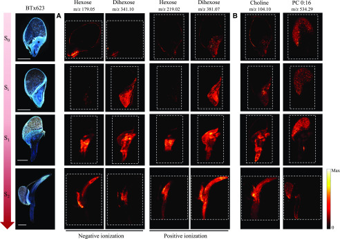
Figure 5.
Distribution and compartmentalization of general metabolites during germination and early seedling development of the cv BTx623 wild-type line. Three consecutive cryo-sections were imaged: The optical images on the far left (cv BTx623) were acquired with fluorescence microscopy, and the remaining images using MALDI-MSI in both negative and positive ion mode, using respectively DAN and DHB as matrix at 30 µm spatial resolution. Developmental stages are as in Figure 2. The dashed lines define the outlines of the recorded MS images. A, Comparative distribution of hexose and dihexose using the two ionization modes. In positive mode, compounds were detected as potassium adducts ([M+K]+), and in negative mode as deprotonated ions ([M-H]−). B, Specific tissue compartmentalization of choline [M+H]+ and phosphocholine (PC 0:16) [M+K]+. Scale bar = 1 mm. Signals for all MSI images are normalized to TIC on each pixel, and maximum values for generating images are listed in Supplemental Figure S4.

