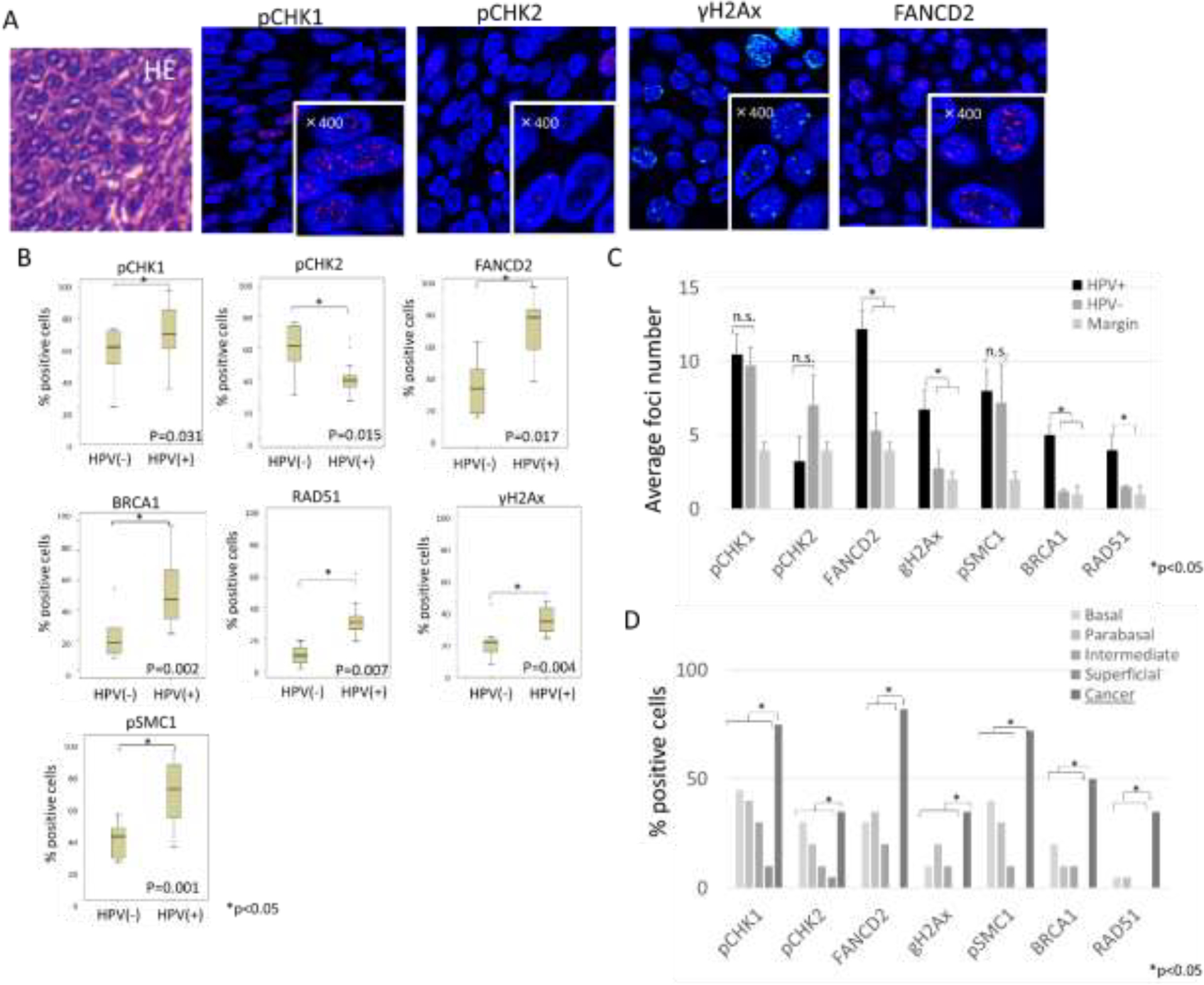
Fig. 2. Expression patterns of DNA damage factors in tumor specimens.

(A) Representative example of HE staining and Immunofluorescence staining of DNA damage factors in HPV positive cancer region. High magnification (x400) is shown in lower right corner and indicates the presence of positive foci. (B) Comparative analysis of the percentages of cells that exhibited any focal localization of DDR factors between HPV positive and negative OPSCC samples. Error bars represent the SD between samples. *P <0.05. (C) Quantitation of average nuclear foci number per positive cell. (D) Comparative analysis of the percentages of cells that exhibited any focal localization of DDR factors between HPV positive OPSCC and normal regions of epithelia in margin (20 or more cells per group were examined).
