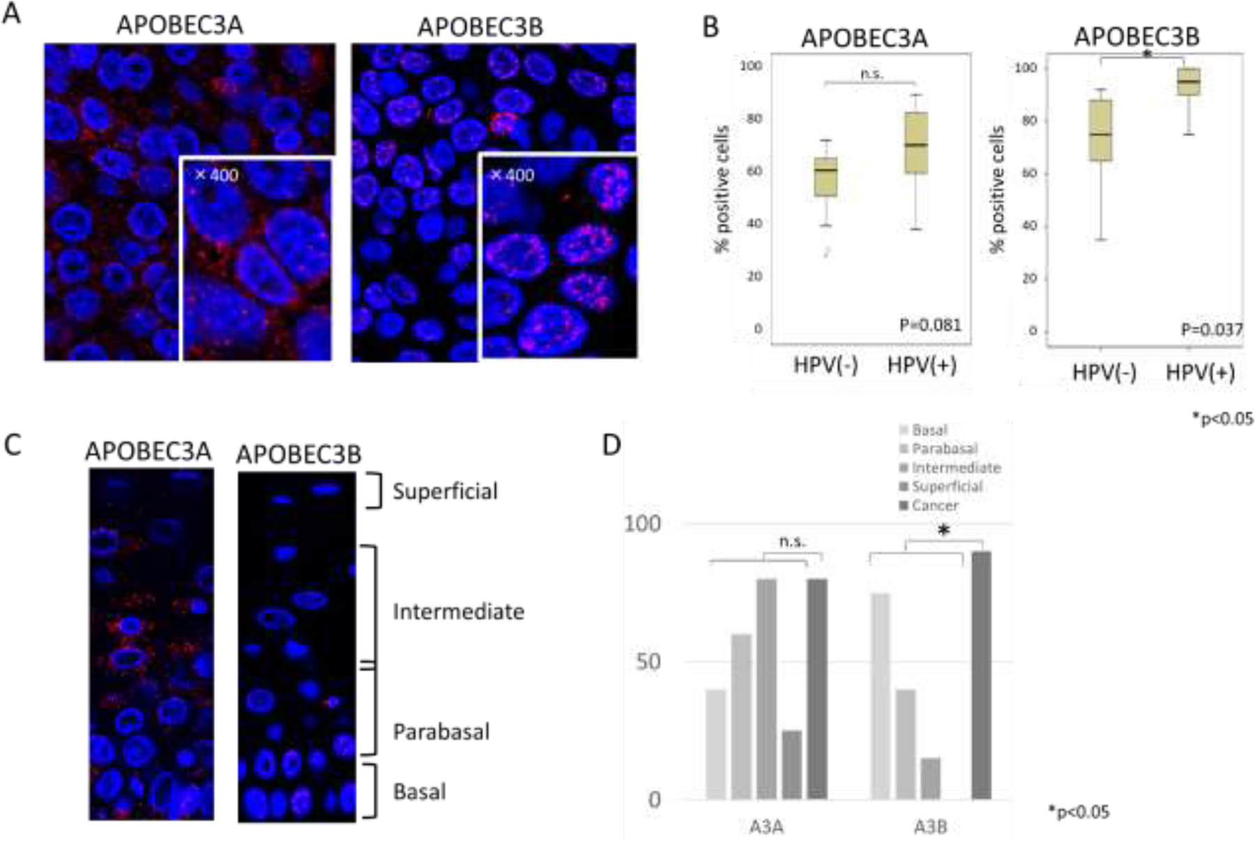
Fig. 3. Expression patterns of APOBEC3A and 3B in tumor specimens.

(A) Representative immunofluorescence staining of APOBEC3A and 3B in HPV positive tumor samples. High magnification (x400) are shown in lower right corner. APOBEC3A antibody used in this analysis was from Sigma while APOBEC3B antibody was from Novus.
(B) Comparative analysis of the percentages of cells that exhibited any focal localization of APOBEC3A and 3B between HPV positive and negative OPSCC samples.
(C) Immunofluorescence staining of APOBEC3A and 3B in normal epithelial layers
(D) Comparative analysis of the percentages of cells that exhibited any focal localization of APOBEC3A and 3B between HPV positive OPSCC and normal epithelia (20 or more cells per group were examined). (P value; 0.17 (APOBEC3A), 0.01(APOBEC3B))
