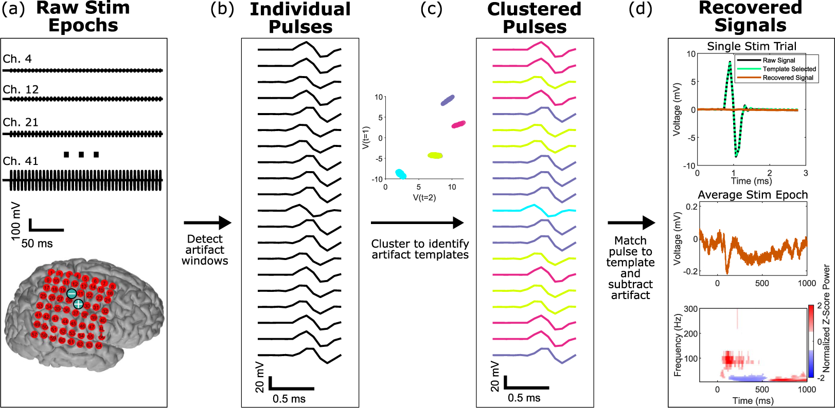
Figure 1.

Schematic overview of our method for signal recovery with stimulation artifacts. (a) Raw stimulation signal epochs (time × channel × epoch) are recorded across an array of electrodes, as shown on a cortical reconstruction of one patient. The two electrode locations indicated by blue ⊕ and ⊖ signs were the sites of the electrical stimulation. These are the input for our algorithm. (b) Individual pulses are identified and extracted within each of these stimulation epoch time periods across all the channels in the array. A small random subset are visualized here. (c) An unsupervised hierarchical density-based clustering technique (HDBSCAN) is used to cluster the individual pulses. Each pulse is colored by the artifact template to which it clustered.(d) Signals are recovered by subtraction of the closest artifact template for each pulse. Subsequent analyses can then be performed directly on the output signals, which are the same size as the input data.
