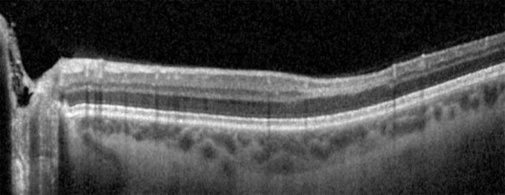
Fig. 1. Optical coherence tomography image from patient 1.

Focal area of hyper-reflective change in the inner and outer plexiform layers with inner nuclear layer volume loss consistent with paracentral acute middle maculopathy.

Focal area of hyper-reflective change in the inner and outer plexiform layers with inner nuclear layer volume loss consistent with paracentral acute middle maculopathy.