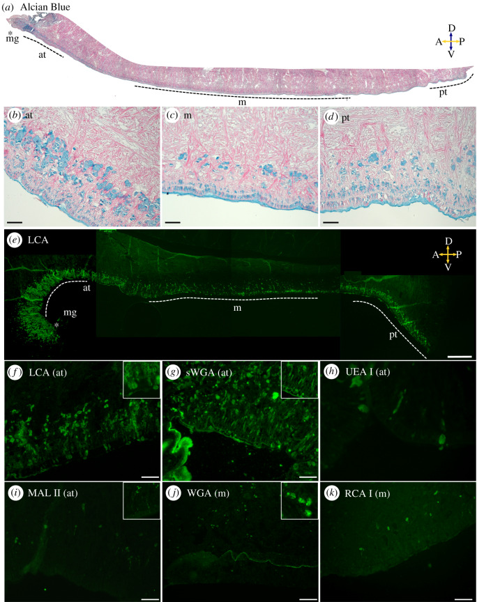
Figure 4.
Overview of P. vulgata foot tissue and the chemistry of glandular secretions. (a–d) Alcian blue (pH 2.5) highlights glands in blue (carboxylate and sulfate moieties) and phloxine stains muscles in red. Regions used for higher-magnification images (b–d) are labelled as marginal groove (mg), anterior (at), middle (m) and posterior (pt). Scale bars: 100 µm in (b–d). Compass labels: A, anterior; P, posterior; D, dorsal; V, ventral. (e–k) Lectin stains highlight the different sugar residues present within specific glands. (e) LCA: stitched image showing the entire foot. Glands contain α-Man and/or α-Fuc linked to N-acetylchitobiose sugar residues. Side-wall glands are not stained. Dotted line marks the epithelium, and imaged regions are labelled as in (a). Scale bar 400 µm. (f) LCA (at): stained glands are found within the epithelium and up to approximately 300 µm into the foot. Scale bar 100 µm. Stained mucus and gland secretions are visible (inset, 150 µm box). (g) sWGA (at): secretions containing GlcNAc but not sialic acid are highlighted throughout the tissue. Scale bar 100 µm. Granules are secreted from long necks (inset, 200 µm box). (h) UEA I (at): specific glands contain α-Fuc. Scale bar 20 µm. (i) MAL II (at): infrequent glands deep in the tissue (approx. 100–150 µm) contain (α-2,3)-sialic acid. Scale bar 100 µm. Granules are secreted from long necks (inset, 370 µm box). (j) WGA (m): glands with GlcNAc are present throughout the foot tissue. Note the epithelium is strongly stained. Glands approximately 30 µm in size are full of granules (inset, 150 µm box). (k) RCA I (m): similar to MAL II but with GalNAc or galactose. Scale bar 50 µm. See text and table 1 for additional information on the lectins used and their ligands.

