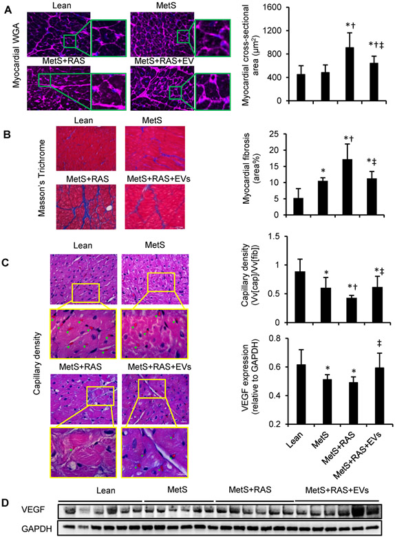
Figure 3.
Extracellular vesicles (EVs) alleviated myocardial remodeling and fibrosis. (A) Left-ventricular sections stained with wheat germ agglutinin (WGA) and quantification of myocyte cross-sectional area. (B) Representative staining and quantification of Masson’s Trichrome. n=6/group. (C) Representative LV sections stained with hematoxylin and eosin, showing capillaries (green arrowheads) and density quantification. (D) Western blotting of vascular endothelial growth-factor (VEGF) in LV. n=6/group. *p<0.05 vs. Lean, †p<0.05 vs. MetS, ‡p<0.05 vs. MetS+RAS. Scale bar=50μm.

