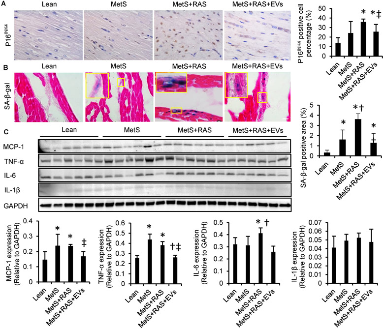
Figure 4.
Extracellular vesicles (EVs) ameliorated myocardial cellular senescence-like phenotype and attenuated myocardial inflammation. (A) Representative immunohistochemical left-ventricular (LV) staining with p16INK4 and quantification. (B) LV sections stained with senescence-associated beta-galactosidase (SA-ß-gal, [blue]) and eosin (red) at pH 6, and quantification. n=6/group. (C) Western blotting of LV monocyte-chemoattractant protein (MCP)-1, tumor necrosis-factor (TNF)-a, interleukin (IL)-6, and IL-1ß and quantification. n=6/group. *p<0.05 vs. Lean, †p<0.05 vs. MetS, ‡p<0.05 vs. MetS+RAS. Scale bar=50μm.

