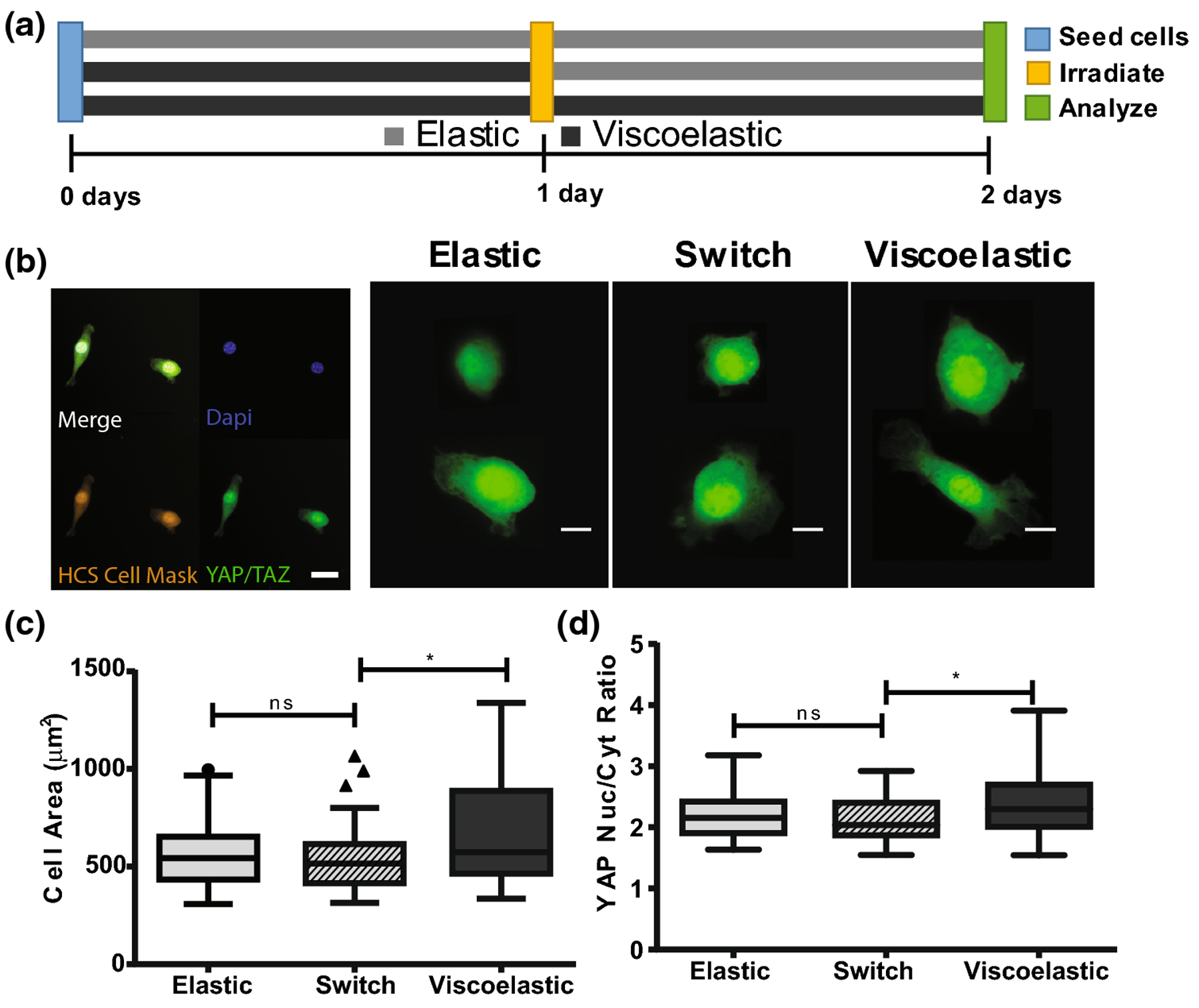
FIGURE 5.

(a) NIH 3T3 fibroblasts were seeded onto swollen 2D thioester hydrogels and cultured for 48 hours. Cells were seeded onto an elastic thioester condition, a viscoelastic thioester condition and a switch condition where viscoelastic hydrogels were made elastic after 24 hours of culture. (b) Cells were fixed at 48 hours and stained for HCS Cell Mask (orange), DAPI (blue) and YAP/TAZ (green); pictured scalebar is 30 microns. YAP/TAZ staining for select cells is shown; the red circle indicates the outline of the nucleus as identified by DAPI staining. Scale bar for single cell images is 10 microns. (c) Spread cell area is visualized by a Tukey boxplot. (d) Nuclear to cytoplasmic ratio of YAP. Reported statistics were calculated using a one tailed student’s t-test, and a population of n=50–60 cells for each condition. Greater than 40 cells were quantified per hydrogel, then 10 cells were selected at random and added to the final population sampling for that condition. (*p<0.05, **p<0.01, ns nonsignificant)
