
Figure 3. Neuronal Differentiation and Self-organizing in HMOs.
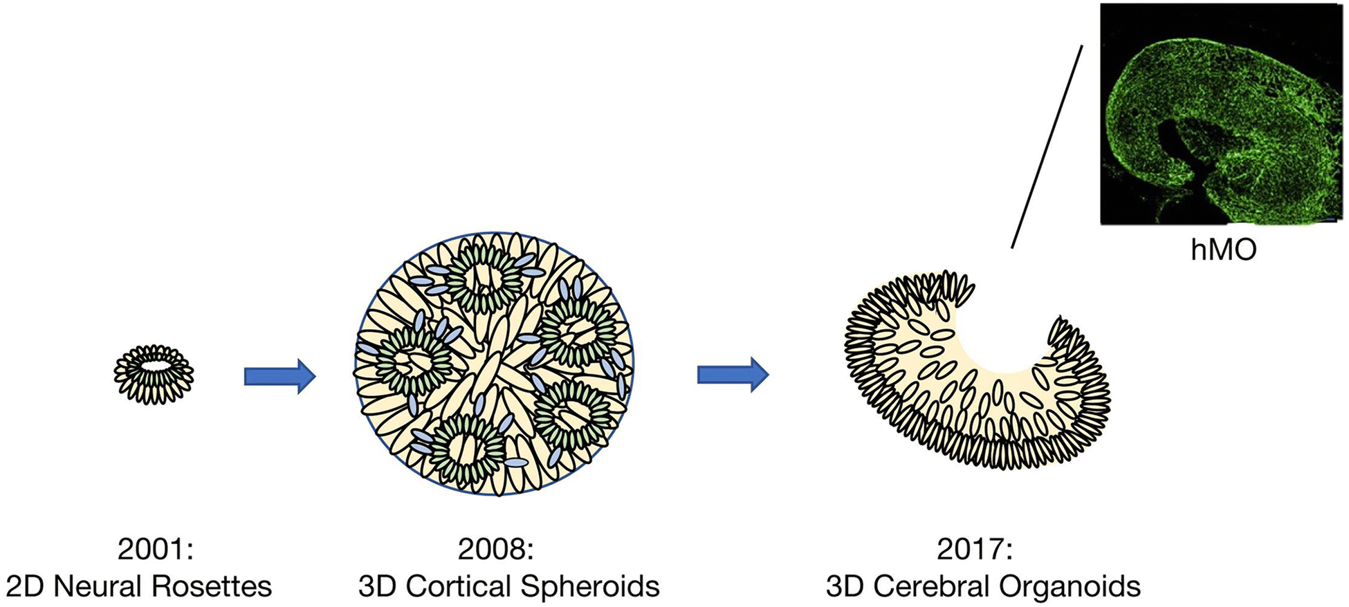
Starting from the initial development of neural rosettes in 2001, developmental models of the brain have become more complex and representative of corticogenesis. In 2008, the development of cortical spheroids allowed for the modelling of both deep and superficial cortical neurons, which then self-organized in a manner that resembled early corticogenesis. More recently, human pluripotent stem cell (hPSC) derived cerebral organoids allow for self-organization and self-patterning of several brain regions. Cerebral organoids have also enabled the development of more specialized structures, such as human midbrain-specific organoids (hMOs). Immunostaining for midbrain dopaminergic neuron (mDN) markers TUJ1 (green) reveals clearly specified clusters of mDNs within hMOs124*. In the schematic, green represents neural stem cells, cream represents intermediate progenitors and blue represents migrating or mature neurons.
*This work is licensed under the Creative Commons Attribution-NonCommercial-NoDerivatives 4.0 International License. To view a copy of this license, visit http://creativecommons.org/licenses/by-ncnd/4.0/ or send a letter to Creative Commons, PO Box 1866, Mountain View, CA 94042, USA. .US