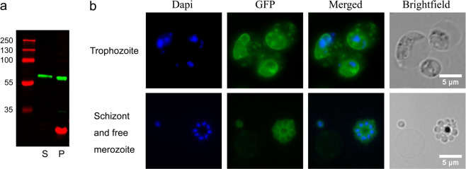
Fig. 5. GFP fusion protein is expressed in transfected parasites.
Protein expression analysis of PYYM_0017400.GFP knock-in cell line. a For Western blot analysis trophozoite-stage parasites were enriched by centrifugation on a Histodenz gradient. Cells were subsequently lysed using saponin resulting in a parasite (P) and supernatant (S) fraction. Exported protein is expected to be seen in the S fraction. A protein of size 63 kDa which correspond to the size of the full-length PYYM_0017400.GFP protein is detected in both supernatant and pellet fraction. The red band corresponds to EXP2 which is a parasitophorous vacoule protein used as a control for the lysis. b Live trophozoites observed under florescence microscopy showed PYYM_0017400.GFP localizing in the PVM and in punctate dots. PYYM_0017400.GFP is found to surround individual merozoites in late schizonts and free merozoites. DNA was stained using Hoechst33342.

